


Journal of Computer Applications ›› 2023, Vol. 43 ›› Issue (4): 1278-1283.DOI: 10.11772/j.issn.1001-9081.2022030478
• Multimedia computing and computer simulation • Previous Articles
Yue CHI, Zhengping LI( ), Chao XU, Bo FENG
Received:2022-04-13
Revised:2022-09-09
Accepted:2022-09-14
Online:2023-01-11
Published:2023-04-10
Contact:
Zhengping LI
About author:CHI Yue, born in 1996, M. S. candidate. His research interests include image processing.Supported by:通讯作者:
李正平
作者简介:池月(1996—),男,河南南阳人,硕士研究生,主要研究方向:图像处理;基金资助:CLC Number:
Yue CHI, Zhengping LI, Chao XU, Bo FENG. Highlight removal algorithm for medical endoscopic images[J]. Journal of Computer Applications, 2023, 43(4): 1278-1283.
池月, 李正平, 徐超, 冯博. 医用内窥镜图像的高光移除算法[J]. 《计算机应用》唯一官方网站, 2023, 43(4): 1278-1283.
Add to citation manager EndNote|Ris|BibTeX
URL: http://www.joca.cn/EN/10.11772/j.issn.1001-9081.2022030478

Fig. 1 Flow of highlight localization
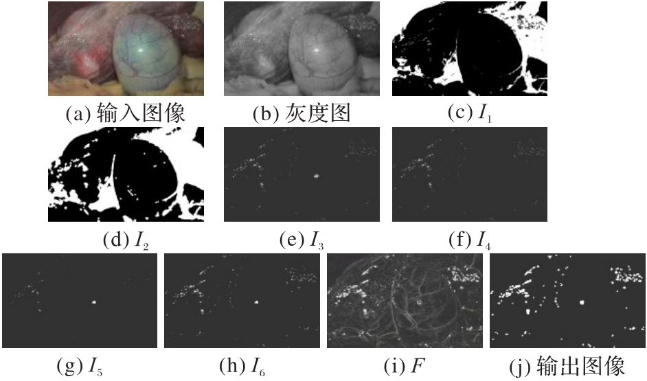
Fig. 2 Example picture

Fig. 3 Flows of original Criminisi algorithm and the proposed algorithm
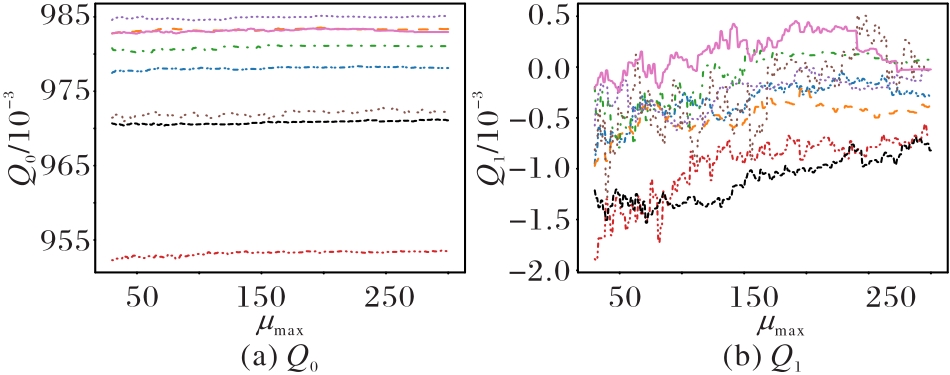
Fig. 4 Line charts of Q0 and Q1 changing with μmax

Fig. 5 Influence of different size templates on effect of vascular reconstruction
| 算法 | 食道1 | 食道2 | 咽部 | 腹腔1 | 腹腔2 | 腹腔3 | 腹腔4 | 鼻部 | 子宫1 | 子宫2 |
|---|---|---|---|---|---|---|---|---|---|---|
| 原图 | 7.992 8 | 5.261 3 | 7.111 6 | 6.926 3 | 6.950 3 | 6.443 1 | 5.959 0 | 7.235 7 | 3.699 5 | 6.765 2 |
| 双色反射模型[ | 7.861 9 | 5.200 5 | 7.060 5 | 6.761 9 | 6.585 0 | 6.463 5 | 6.885 3 | 7.606 7 | 3.763 9 | 6.735 7 |
| RPCA[ | 7.162 1 | 5.083 3 | 6.617 5 | 6.614 4 | 6.378 8 | 5.829 7 | 6.647 8 | 7.253 3 | 3.771 7 | 6.733 9 |
| 热扩散[ | 6.908 7 | 5.120 8 | 6.453 6 | 6.705 3 | 6.359 0 | 5.967 9 | 5.610 7 | 7.350 5 | 3.868 9 | 6.757 9 |
| Criminisi[ | 6.543 3 | 5.092 7 | 6.282 5 | 6.606 0 | 6.128 1 | 5.654 0 | 5.795 3 | 7.233 5 | 3.745 3 | 6.884 0 |
| 本文算法 | 6.164 2 | 5.032 3 | 5.931 5 | 6.581 8 | 5.514 6 | 5.464 2 | 5.535 0 | 7.204 5 | 3.683 9 | 6.723 7 |
Tab. 1 NIQE test results of 10 images under different algorithms
| 算法 | 食道1 | 食道2 | 咽部 | 腹腔1 | 腹腔2 | 腹腔3 | 腹腔4 | 鼻部 | 子宫1 | 子宫2 |
|---|---|---|---|---|---|---|---|---|---|---|
| 原图 | 7.992 8 | 5.261 3 | 7.111 6 | 6.926 3 | 6.950 3 | 6.443 1 | 5.959 0 | 7.235 7 | 3.699 5 | 6.765 2 |
| 双色反射模型[ | 7.861 9 | 5.200 5 | 7.060 5 | 6.761 9 | 6.585 0 | 6.463 5 | 6.885 3 | 7.606 7 | 3.763 9 | 6.735 7 |
| RPCA[ | 7.162 1 | 5.083 3 | 6.617 5 | 6.614 4 | 6.378 8 | 5.829 7 | 6.647 8 | 7.253 3 | 3.771 7 | 6.733 9 |
| 热扩散[ | 6.908 7 | 5.120 8 | 6.453 6 | 6.705 3 | 6.359 0 | 5.967 9 | 5.610 7 | 7.350 5 | 3.868 9 | 6.757 9 |
| Criminisi[ | 6.543 3 | 5.092 7 | 6.282 5 | 6.606 0 | 6.128 1 | 5.654 0 | 5.795 3 | 7.233 5 | 3.745 3 | 6.884 0 |
| 本文算法 | 6.164 2 | 5.032 3 | 5.931 5 | 6.581 8 | 5.514 6 | 5.464 2 | 5.535 0 | 7.204 5 | 3.683 9 | 6.723 7 |
| 样图 | 分辨率 | 高光 占比/% | 运行时间/s | ||
|---|---|---|---|---|---|
| RPCA[ | Criminisi[ | 本文算法 | |||
| picture1 | 863×1 027 | 2.60 | 42.657 | 1 755.800 | 21.072 |
| picture2 | 626×721 | 2.54 | 18.022 | 434.800 | 11.531 |
| picture3 | 823×1 062 | 1.89 | 33.131 | 1 303.080 | 13.314 |
| picture4 | 343×338 | 1.21 | 3.207 | 15.741 | 2.271 |
| picture5 | 422×527 | 1.66 | 7.864 | 82.974 | 4.151 |
| picture6 | 847×1 001 | 3.67 | 33.067 | 2 142.800 | 30.423 |
| picture7 | 492×475 | 2.31 | 8.349 | 112.633 | 5.769 |
| picture8 | 527×549 | 2.26 | 12.240 | 88.946 | 11.478 |
Tab. 2 Comparison of runtime of different algorithms
| 样图 | 分辨率 | 高光 占比/% | 运行时间/s | ||
|---|---|---|---|---|---|
| RPCA[ | Criminisi[ | 本文算法 | |||
| picture1 | 863×1 027 | 2.60 | 42.657 | 1 755.800 | 21.072 |
| picture2 | 626×721 | 2.54 | 18.022 | 434.800 | 11.531 |
| picture3 | 823×1 062 | 1.89 | 33.131 | 1 303.080 | 13.314 |
| picture4 | 343×338 | 1.21 | 3.207 | 15.741 | 2.271 |
| picture5 | 422×527 | 1.66 | 7.864 | 82.974 | 4.151 |
| picture6 | 847×1 001 | 3.67 | 33.067 | 2 142.800 | 30.423 |
| picture7 | 492×475 | 2.31 | 8.349 | 112.633 | 5.769 |
| picture8 | 527×549 | 2.26 | 12.240 | 88.946 | 11.478 |

Fig. 6 HSV channel localization effect

Fig. 7 Highlight localization effect of different methods

Fig. 8 Effect of different highlight removal algorithms
| 1 | MORO C, ŠTROMBERGA Z, RAIKOS A, et al. The effectiveness of virtual and augmented reality in health sciences and medical anatomy[J]. Anatomical Sciences Education, 2017, 10(6): 549-559. 10.1002/ase.1696 |
| 2 | BERNHARDT S, NICOLAU S A, SOLER L, et al. The status of augmented reality in laparoscopic surgery as of 2016[J]. Medical Image Analysis, 2017, 37: 66-90. 10.1016/j.media.2017.01.007 |
| 3 | BUCHS N C, VOLONTÉ F, PUGIN F, et al. Augmented environments for the targeting of hepatic lesions during image-guided robotic liver surgery[J]. Journal of Surgical Research, 2013, 184(2): 825-831. 10.1016/j.jss.2013.04.032 |
| 4 | WHITE S, KALKOFEN D, SANDOR C. Visualization in mixed reality environments[C]// Proceedings of the 2011 IEEE International Symposium on Mixed and Augmented Reality - Arts, Media and Humanities. Piscataway: IEEE, 2011: 1-1. 10.1109/ismar.2011.6092361 |
| 5 | KERSTEN-OERTEL M, JANNIN P, COLLINS D L. The state of the art of visualization in mixed reality image guided surgery[J]. Computerized Medical Imaging and Graphics, 2013, 37(2): 98-112. 10.1016/j.compmedimag.2013.01.009 |
| 6 | OH J, HWANG S, LEE J, et al. Informative frame classification for endoscopy video[J]. Medical Image Analysis, 2007, 11(2): 110-127. 10.1016/j.media.2006.10.003 |
| 7 | 卢桂荣,汤景凡,姜明. 基于快速双边滤波的图像高光去除研究[J]. 计算机工程与应用, 2014, 50(10): 176-179, 207. 10.3778/j.issn.1002-8331.1310-0359 |
| LU G R, TANG J F, JIANG M. Research on image highlight removal based on fast bilateral filtering[J]. Computer Engineering and Applications, 2014, 50(10): 176-179, 207. 10.3778/j.issn.1002-8331.1310-0359 | |
| 8 | REN W H, TIAN J D, TANG Y D. Specular reflection separation with color-lines constraint[J]. IEEE Transactions on Image Processing. 2017, 26(5): 2327-2337. 10.1109/tip.2017.2675204 |
| 9 | YANG Q X, TANG J H, AHUJA N. Efficient and robust specular highlight removal[J]. IEEE Transactions on Pattern Analysis and Machine Intelligence, 2015, 37(6): 1304-1311. 10.1109/tpami.2014.2360402 |
| 10 | SHEN H L, ZHANG H G, SHAO S J, et al. Chromaticity-based separation of reflection components in a single image[J]. Pattern Recognition, 2008, 41(8):2461-2469. 10.1016/j.patcog.2008.01.026 |
| 11 | KHAN H A, THOMAS J B, HARDEBERG J Y. Analytical survey of highlight detection in color and spectral images[C]// Proceedings of the 2017 International Workshop on Computational Color Imaging, LNCS 10213. Cham: Springer, 2017:197-208. |
| 12 | JOSPIN L V, BAECHLER G, SCHOLEFIELD A. Embedded polarizing filters to separate diffuse and specular reflection[C]// Proceedings of the 2018 Asian Conference on Computer Vision, LNCS 11362. Cham: Springer, 2019:3-18. |
| 13 | SUO J L, AN D S, JI X Y, et al. Fast and high quality highlight removal from a single image[J]. IEEE Transactions on Image Processing, 2016, 25(11): 5441-5454. 10.1109/tip.2016.2605002 |
| 14 | SHEN H L, ZHENG Z H. Real-time highlight removal using intensity ratio[J]. Applied Optics, 2013, 52(19): 4483-4493. 10.1364/ao.52.004483 |
| 15 | LI R Y, PAN J J, SI Y Q, et al. Specular reflections removal for endoscopic image sequences with adaptive-RPCA decomposition[J]. IEEE Transactions on Medical Imaging, 2020, 39(2): 328-340. 10.1109/tmi.2019.2926501 |
| 16 | BOBROW T L, MAHMOOD F, INSERNI M, et al. DeepLSR: a deep learning approach for laser speckle reduction[J]. Biomedical Optics Express, 2019, 10(6):2869-2882. 10.1364/boe.10.002869 |
| 17 | KUDVA V, PRASAD K, GURUVARE S. Detection of specular reflection and segmentation of cervix region in uterine cervix images for cervical cancer screening[J]. IRBM, 2017, 38(5): 281-291. 10.1016/j.irbm.2017.08.003 |
| 18 | SAINT-PIERRE C A, BOISVERT J, GRIMARD G, et al. Detection and correction of specular reflections for automatic surgical tool segmentation in thoracoscopic images[J]. Machine Vision and Applications, 2011, 22(1): 171-180. 10.1007/s00138-007-0099-6 |
| 19 | LIANG Z S, YANG G B, DING X L, et al. An efficient forgery detection algorithm for object removal by exemplar-based image inpainting[J]. Journal of Visual Communication and Image Representation, 2015, 30: 75-85. 10.1016/j.jvcir.2015.03.004 |
| 20 | QURESHI M A, DERICHE M, BEGHDADI A, et al. A critical survey of state-of-the-art image inpainting quality assessment metrics[J]. Journal of Visual Communication and Image Representation, 2017, 49: 177-191. 10.1016/j.jvcir.2017.09.006 |
| 21 | BERTALMIO M, BERTOZZI A L, SAPIRO G. Navier-stokes, fluid dynamics, and image and video inpainting[C]// Proceedings of the 2001 IEEE Computer Society Conference on Computer Vision and Pattern Recognition. Piscataway: IEEE, 2001: I-355-I-362. 10.1109/cvpr.2001.990916 |
| 22 | CRIMINISI A, PÉREZ P, TOYAMA K. Region filling and object removal by exemplar-based image inpainting[J]. IEEE Transactions on Image Processing, 2004, 13(9): 1200-1212. 10.1109/tip.2004.833105 |
| 23 | BUYSSENS P, DAISY M, TSCHUMPERLÉ D, et al. Exemplar-based inpainting: technical review and new heuristics for better geometric reconstructions[J]. IEEE Transactions on Image Processing, 2015, 24(6): 1809-1824. |
| 24 | 王凤随,刘正男,付林军. 基于信息熵和梯度因子的改进Criminisi图像修复方法[J]. 激光与光电子学进展, 2020, 57(22): No.221006. 10.3788/lop57.221006 |
| WANG F S, LIU Z N, FU L J. An improved Criminisi image inpainting method based on information entropy and gradient factor[J]. Progress in Lasers and Optoelectronics, 2020, 57(22): No.221006. 10.3788/lop57.221006 | |
| 25 | KUMAR S, SRIVASTAVA A K, JHA A, et al. Implementation of linear structuring element in OpenCV for blood vessel segmentation from color fundus images[C]// Proceedings of the 10th International Conference on Computing, Communication and Networking Technologies. Piscataway: IEEE, 2019:1-5. 10.1109/icccnt45670.2019.8944610 |
| 26 | MITTAL A, SOUNDARARAJIAN R, BOVIK A C. Making a “completely blind” image quality analyzer[J]. IEEE Signal Processing Letters, 2013, 20(3): 209-212. 10.1109/lsp.2012.2227726 |
| 27 | TELEA A. An image inpainting technique based on the fast marching method[J]. Journal of Graphics Tools, 2004, 9(1): 23-34. 10.1080/10867651.2004.10487596 |
| [1] | WEI Zhenyu, WEN Chang, XIE Kai, HE Jianbiao. Real-time face detection for mobile devices with optical flow estimation [J]. Journal of Computer Applications, 2018, 38(4): 1146-1150. |
| [2] | LI Mengxue, ZHAI Donghai, MENG Hongyue, CAO Daming. Image inpainting algorithm for partitioning feature subregions [J]. Journal of Computer Applications, 2017, 37(12): 3541-3546. |
| [3] | ZHAI Donghai XIAO Jie YU Jiang LI Tongliang. Image inpainting algorithm based on adaptive template [J]. Journal of Computer Applications, 2013, 33(10): 2891-2894. |
| [4] | Hai-yang WANG Sheng-bing CHE Xu SHU. Semi-fragile watermarking algorithm based on dynamic image segmentation and information entropy [J]. Journal of Computer Applications, 2011, 31(08): 2169-2173. |
| [5] | . Multi-focus image fusion algorithm based on region segmentation and nonsubsampled Contourlet transform [J]. Journal of Computer Applications, 2010, 30(10): 2805-2807. |
| [6] | Qiang-Feng ZHOU Zheng Tian Xiao-bin LI Bing-Tao LIU. Effective implementation of image region segmentation based on Gomory-Hu algorithm [J]. Journal of Computer Applications, 2008, 28(3): 671-673. |
| Viewed | ||||||
|
Full text |
|
|||||
|
Abstract |
|
|||||