


《计算机应用》唯一官方网站 ›› 2025, Vol. 45 ›› Issue (6): 1998-2006.DOI: 10.11772/j.issn.1001-9081.2024060855
蒋杰1, 骆功宁1, 董素宇2, 李凡丁1, 李向宇1( ), 李钦策1, 袁永峰1, 王宽全1
收稿日期:2024-06-24
修回日期:2024-09-06
接受日期:2024-09-10
发布日期:2024-09-18
出版日期:2025-06-10
通讯作者:
李向宇
作者简介:蒋杰(2001—),男,重庆人,硕士研究生,主要研究方向:医学图像处理、计算机视觉
Jie JIANG1, Gongning LUO1, Suyu DONG2, Fanding LI1, Xiangyu LI1(), Qince LI1, Yongfeng YUAN1, Kuanquan WANG1
Received:2024-06-24
Revised:2024-09-06
Accepted:2024-09-10
Online:2024-09-18
Published:2025-06-10
Contact:
Xiangyu LI
About author:JIANG Jie, born in 2001, M. S. candidate. His research interests include medical image processing, computer vision.Supported by:摘要:
在计算机辅助诊断领域,精确分割计算机断层扫描(CT)图像中的颅内出血(ICH)对后续的治疗和预后至关重要。针对小出血区域难以分割的问题,提出一种信息瓶颈引导的ICH分割方法并基于该方法构建一个信息瓶颈引导的分割网络(IBGS-Net)。首先,采用U-Net架构作为基础,并引入信息瓶颈层增强与ICH分割相关的关键特征的识别;其次,通过设计的残差空间自适应归一化(ResSPADE)模块,信息激活图(IAM)被有效整合到分割流程中,提升网络对出血区域的识别和定位能力;最后,引入交互引导损失(IGL)函数以优化模型对难分割区域的处理,进一步增强模型的泛化性能。在内部数据集上的评估结果表明,所提方法在Dice相似性系数(DSC)、归一化表面Dice(NSD)和相对体积差(RVD)这3个指标上分别达到了78.1%、90.1%和11.5%;在公开数据集INSTANCE 2022上,与其他的分割方法的比较结果表明,所提方法的3个指标相较于次优结果,分别提升了1.9、2.4和下降了3.2个百分点。以上验证了所提方法在ICH分割任务中的有效性和优越性,可用于协助临床医生进行ICH分割。
中图分类号:
蒋杰, 骆功宁, 董素宇, 李凡丁, 李向宇, 李钦策, 袁永峰, 王宽全. 信息瓶颈引导的颅内出血分割方法[J]. 计算机应用, 2025, 45(6): 1998-2006.
Jie JIANG, Gongning LUO, Suyu DONG, Fanding LI, Xiangyu LI, Qince LI, Yongfeng YUAN, Kuanquan WANG. Information bottleneck-guided intracranial hemorrhage segmentation method[J]. Journal of Computer Applications, 2025, 45(6): 1998-2006.
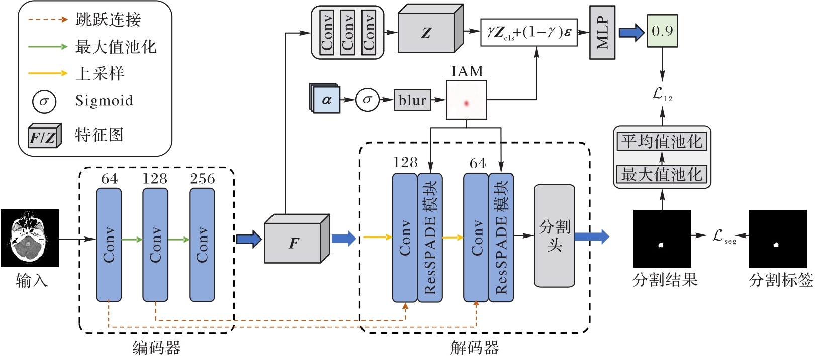
图1 本文模型的结构
Fig. 1 Structure of proposed model
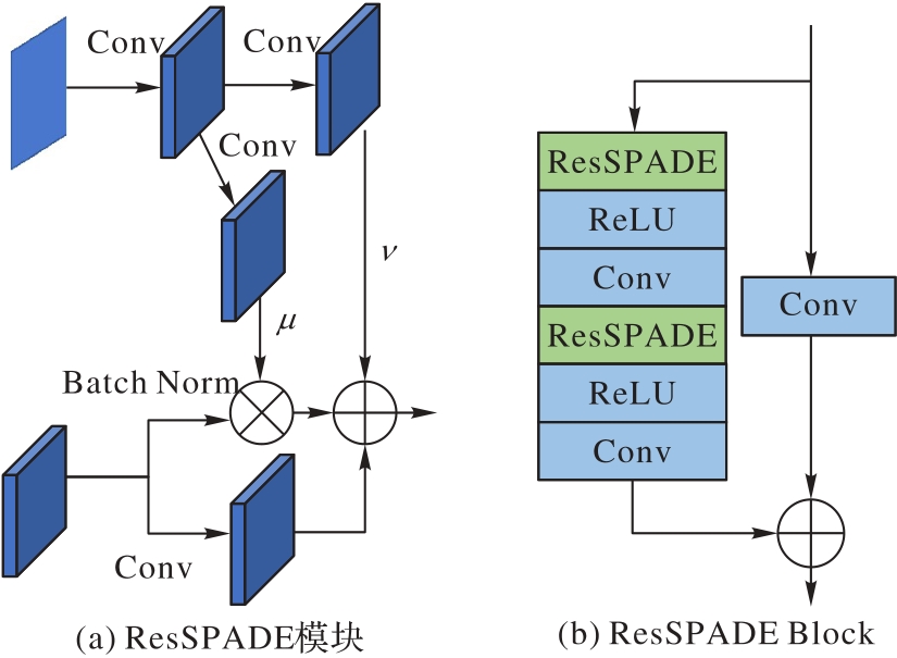
图2 ResSPADE结构和ResSPADE Block结构
Fig.2 ResSPADE structure and ResSPADE Block structure
| 数据集 | 方法 | Dice | NSD | RVD |
|---|---|---|---|---|
| 内部数据集 | Patch-FCN[ | 75.5 | 84.9 | 17.2 |
| DR-UNet[ | 76.3 | 85.4 | 14.3 | |
| U-Net[ | 75.8 | 83.9 | 21.2 | |
| 3D U-Net[ | 76.4 | 87.0 | 16.2 | |
| SLEX-Net[ | 76.1 | 86.8 | 15.7 | |
| IBGS-Net | 78.1 | 90.1 | 11.5 | |
| 公共数据集 | UNETR[ | 60.7 | 45.4 | 47.8 |
| U-Net[ | 57.5 | 41.5 | 54.3 | |
| 3D U-Net[ | 59.3 | 46.3 | 36.3 | |
| CHSNet[ | 61.3 | 45.2 | 35.2 | |
| MOEL-Net[ | 58.9 | 43.4 | 40.3 | |
| IBGS-Net | 63.2 | 48.7 | 32.0 |
表1 不同方法的分割性能对比 (%)
Tab. 1 Comparison of segmentation performance of different methods
| 数据集 | 方法 | Dice | NSD | RVD |
|---|---|---|---|---|
| 内部数据集 | Patch-FCN[ | 75.5 | 84.9 | 17.2 |
| DR-UNet[ | 76.3 | 85.4 | 14.3 | |
| U-Net[ | 75.8 | 83.9 | 21.2 | |
| 3D U-Net[ | 76.4 | 87.0 | 16.2 | |
| SLEX-Net[ | 76.1 | 86.8 | 15.7 | |
| IBGS-Net | 78.1 | 90.1 | 11.5 | |
| 公共数据集 | UNETR[ | 60.7 | 45.4 | 47.8 |
| U-Net[ | 57.5 | 41.5 | 54.3 | |
| 3D U-Net[ | 59.3 | 46.3 | 36.3 | |
| CHSNet[ | 61.3 | 45.2 | 35.2 | |
| MOEL-Net[ | 58.9 | 43.4 | 40.3 | |
| IBGS-Net | 63.2 | 48.7 | 32.0 |
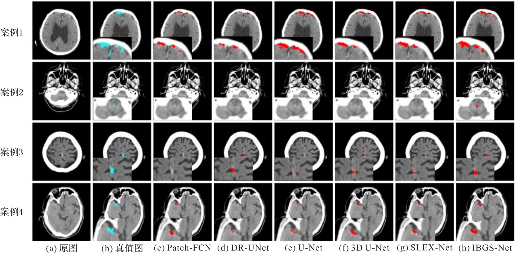
图3 不同分割方法结果的可视化
Fig. 3 Visualization of results of different segmentation methods
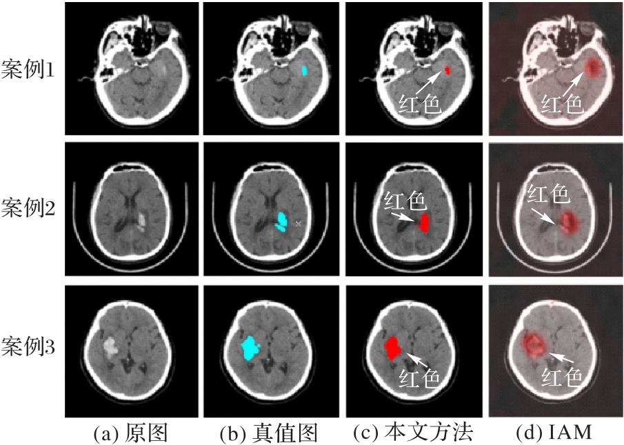
图4 IAM的可视化
Fig. 4 IAM visualization
| 实验 | IAM | ResSPADE | IGL | Dice | NSD | RVD |
|---|---|---|---|---|---|---|
| A | 75.7 | 84.1 | 19.2 | |||
| B | √ | 76.1 | 84.7 | 15.5 | ||
| C | √ | √ | 76.9 | 87.3 | 13.2 | |
| D | √ | 77.2 | 86.9 | 14.1 | ||
| E | √ | √ | √ | 78.1 | 90.1 | 11.5 |
表2 消融实验结果对比 (%)
Tab. 2 Comparison of ablation experimental results
| 实验 | IAM | ResSPADE | IGL | Dice | NSD | RVD |
|---|---|---|---|---|---|---|
| A | 75.7 | 84.1 | 19.2 | |||
| B | √ | 76.1 | 84.7 | 15.5 | ||
| C | √ | √ | 76.9 | 87.3 | 13.2 | |
| D | √ | 77.2 | 86.9 | 14.1 | ||
| E | √ | √ | √ | 78.1 | 90.1 | 11.5 |
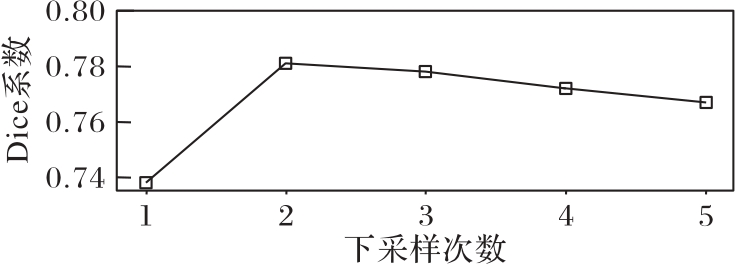
图5 不同下采样次数对分割结果的影响
Fig.5 Influence of different downsampling times on segmentation results
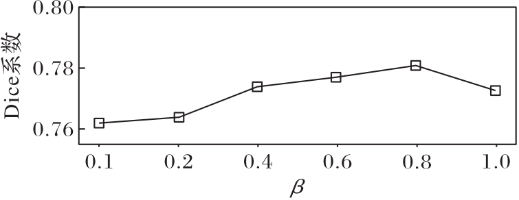
图6 β对ICH分割结果的影响
Fig. 6 Influence of β on segmentation results of ICH
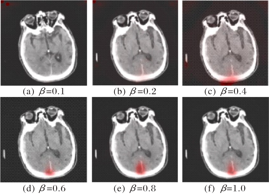
图7 β对ICH病变定位的影响
Fig. 7 Influence of β on localization of ICH lesion
| 1 | VAN ASCH C J J, LUITSE M J A, RINKEL G J E, et al. Incidence, case fatality, and functional outcome of intracerebral haemorrhage over time, according to age, sex, and ethnic origin: a systematic review and meta-analysis[J]. The Lancet Neurology, 2010, 9(2): 167-176. |
| 2 | QURESHI A I, PALESCH Y Y. Antihypertensive Treatment of Acute Cerebral Hemorrhage (ATACH) Ⅱ: design, methods, and rationale [J]. Neurocritical Care, 2011, 15(3): 559-576. |
| 3 | ABRAMOVA V, CLÈRIGUES A, QUILES A, et al. Hemorrhagic stroke lesion segmentation using a 3D U-Net with squeeze-and-excitation blocks [J]. Computerized Medical Imaging and Graphics, 2021, 90: No.101908. |
| 4 | ISLAM M, SANGHANI P, SEE A A Q, et al. ICHNet: IntraCerebral Hemorrhage (ICH) segmentation using deep learning[C]// Proceedings of the 2018 International MICCAI Brainlesion Workshop, LNCS 11383. Cham: Springer, 2019: 456-463. |
| 5 | KOTHARI R U, BROTT T, BRODERICK J P, et al. The ABCs of measuring intracerebral hemorrhage volumes[J]. Stroke, 1996, 27(8): 1304-1305. |
| 6 | WEBB A J S, ULLMAN N L, MORGAN T C, et al. Accuracy of the ABC/2 score for intracerebral hemorrhage: systematic review and analysis of MISTIE, CLEAR-IVH, and CLEAR Ⅲ[J]. Stroke, 2015, 46(9): 2470-2476. |
| 7 | HU Q, QIAN G, AZIZ A, et al. Segmentation of brain from computed tomography head images[C]// Proceedings of the 27th Annual International Conference of the IEEE Engineering in Medicine and Biology Society. Piscataway: IEEE, 2005: 3375-3378. |
| 8 | MAKSIMOVIC R, STANKOVIC S, MILOVANOVIC D. Computed tomography image analyzer: 3D reconstruction and segmentation applying active contour models — ‘snakes’[J]. International Journal of Medical Informatics, 2000, 58/59: 29-37. |
| 9 | RAMTEKE R J, KHACHANE M Y. Automatic medical image classification and abnormality detection using K-nearest neighbour[J]. International Journal of Advanced Computer Research, 2012, 2(6): 190-196. |
| 10 | HSSAYENI M D, CROOCK M S, SALMAN A D, et al. Intracranial hemorrhage segmentation using a deep convolutional model [J]. Data, 2020, 5(1): No.14. |
| 11 | RONNEBERGER O, FISCHER P, BROX T. U-Net: convolutional networks for biomedical image segmentation[C]// Proceedings of the 2015 International Conference on Medical Image Computing and Computer Assisted Intervention, LNCS 9351. Cham: Springer, 2015: 234-241. |
| 12 | CHANG P D, KUOY E, GRINBAND J, et al. Hybrid 3D/2D convolutional neural network for hemorrhage evaluation on head CT[J]. American Journal of Neuroradiology, 2018, 39(9): 1609-1616. |
| 13 | KUANG Z, DENG X, YU L, et al. Ψ-Net: focusing on the border areas of intracerebral hemorrhage on CT images[J]. Computer Methods and Programs in Biomedicine, 2020, 194: No.105546. |
| 14 | LI X, LUO G, WANG W, et al. Hematoma expansion context guided intracranial hemorrhage segmentation and uncertainty estimation[J]. IEEE Journal of Biomedical and Health Informatics, 2022, 26(3): 1140-1151. |
| 15 | SCHULZ K, SIXT L, TOMBARI F, et al. Restricting the flow: information bottlenecks for attribution[EB/OL]. [2024-06-18].. |
| 16 | PARK T, LIU M Y, WANG T C, et al. Semantic image synthesis with spatially-adaptive normalization [C]// Proceedings of the 2019 IEEE/CVF Conference on Computer Vision and Pattern Recognition. Piscataway: IEEE, 2019: 2332-2341. |
| 17 | KWON D, AHN J, KIM J, et al. Siamese U-Net with healthy template for accurate segmentation of intracranial hemorrhage[C]// Proceedings of the 2019 International Conference on Medical Image Computing and Computer Assisted Intervention, LNCS 11766. Cham: Springer, 2019: 848-855. |
| 18 | TOIKKANEN M, KWON D, LEE M. ReSGAN: intracranial hemorrhage segmentation with residuals of synthetic brain CT scans[C]// Proceedings of the 2021 International Conference on Medical Image Computing and Computer Assisted Intervention, LNCS 12901. Cham: Springer, 2021: 400-409. |
| 19 | PIAO Z, GU Y H, JIN H, et al. Intracerebral hemorrhage CT scan image segmentation with HarDNet based transformer[J]. Scientific Reports, 2023, 13: No.7208. |
| 20 | NIJIATI M, TUERSUN A, ZHANG Y, et al. A symmetric prior knowledge based deep learning model for intracerebral hemorrhage lesion segmentation[J]. Frontiers in Physiology, 2022, 13: No.977427. |
| 21 | KYUNG S, SHIN K, JEONG H, et al. Improved performance and robustness of multi-task representation learning with consistency loss between pretexts for intracranial hemorrhage identification in head CT [J]. Medical Image Analysis, 2022, 81: No.102489. |
| 22 | 张鹏,徐曾春,胡平. 融合密集连接与注意机制的颅内出血分割方法[J]. 小型微型计算机系统, 2021, 42(7): 1458-1463. |
| ZHANG P, XU Z C, HU P. Segmentation of intracranial hemorrhage fusing dense connection and attention mechanism[J]. Journal of Chinese Computer Systems, 2021, 42(7): 1458-1463. | |
| 23 | ZHOU B, KHOSLA A, LAPEDRIZA A, et al. Learning deep features for discriminative localization[C]// Proceedings of the 2016 IEEE Conference on Computer Vision and Pattern Recognition. Piscataway: IEEE, 2016: 2921-2929. |
| 24 | SELVARAJU R R, COGSWELL M, DAS A, et al. Grad-CAM: visual explanations from deep networks via gradient-based localization[C]// Proceedings of the 2017 IEEE International Conference on Computer Vision. Piscataway: IEEE, 2017: 618-626. |
| 25 | 张童禹,李恩慧,李振宇, 等. 基于多实例学习及阈值伪标签提取的CT影像颅内出血分割[J]. 中国生物医学工程学报, 2023, 42(6): 677-686. |
| ZHANG T Y, LI E H, LI Z Y, et al. Segmentation of intracranial hemorrhage in CT images based on multi-instance learning and thresholding pseudo-labels extraction[J]. Chinese Journal of Biomedical Engineering, 2023, 42(6):677-686. | |
| 26 | YUN S, HAN D, CHUN S, et al. CutMix: regularization strategy to train strong classifiers with localizable features [C]// Proceedings of the 2019 IEEE/CVF International Conference on Computer Vision. Piscataway: IEEE, 2019: 6022-6031. |
| 27 | BABAR S, DAS S. Where to look?: Mining complementary image regions for weakly supervised object localization[C]// Proceedings of the 2021 IEEE Winter Conference on Applications of Computer Vision. Piscataway: IEEE, 2021: 1009-1018. |
| 28 | KIM J, CHOE J, YUN S, et al. Normalization matters in weakly supervised object localization [C]// Proceedings of the 2021 IEEE/CVF International Conference on Computer Vision. Piscataway: IEEE, 2021: 3407-3416. |
| 29 | BANG S, XIE P, LEE H, et al. Explaining a black-box by using a deep variational information bottleneck approach [C]// Proceedings of the 35th AAAI Conference on Artificial Intelligence. Palo Alto: AAAI Press, 2021: 11396-11404. |
| 30 | SMILKOV D, THORAT N, KIM B, et al. SmoothGrad: removing noise by adding noise [EB/OL]. [2024-06-18].. |
| 31 | KLAMBAUER G, UNTERTHINER T, MAYR A, et al. Self-normalizing neural networks[C]// Proceedings of the 31st International Conference on Neural Information Processing Systems. Red Hook: Curran Associates Inc., 2017: 972-981. |
| 32 | LI X, WANG K, LIU J, et al. The 2022 intracranial hemorrhage segmentation challenge on Non-Contrast head CT (NCCT) (Mar 2022). [EB/OL]. [2024-05-21]. . |
| 33 | LI X, LUO G, WANG K, et al. The state-of-the-art 3D anisotropic intracranial hemorrhage segmentation on non-contrast head CT: the INSTANCE challenge[EB/OL]. [2024-06-18].. |
| 34 | KUO W, HÄNE C, YUH E, et al. Cost-sensitive active learning for intracranial hemorrhage detection[C]// Proceedings of the 2018 International Conference on Medical Image Computing and Computer-Assisted Intervention, LNCS 11072. Cham: Springer, 2018: 715-723. |
| 35 | YU N, YU H, LI H, et al. A robust deep learning segmentation method for hematoma volumetric detection in intracerebral hemorrhage[J]. Stroke, 2022, 53(1): 167-176. |
| 36 | ÇIÇEK Ö, ABDULKADIR A, LIENKAMP S S, et al. 3D U-Net: learning dense volumetric segmentation from sparse annotation[C]// Proceedings of the 2016 International Conference on Medical Image Computing and Computer-Assisted Intervention, LNCS 9901. Cham: Springer, 2016: 424-432. |
| 37 | HATAMIZADEH A, TANG Y, NATH V, et al. UNETR: transformers for 3D medical image segmentation [C]// Proceedings of the 2022 IEEE/CVF Winter Conference on Applications of Computer Vision. Piscataway: IEEE, 2022: 1748-1758. |
| 38 | XU B, FAN Y, LIU J, et al. CHSNet: automatic lesion segmentation network guided by CT image features for acute cerebral hemorrhage[J]. Computers in Biology and Medicine, 2023, 164: No.107334. |
| 39 | ZHANG Y, HUANG Y, HU K. Multi-scale object equalization learning network for intracerebral hemorrhage region segmentation[J]. Neural Networks, 2024, 179: No.106507. |
| [1] | 许立君, 黎辉, 刘祖阳, 陈侃松, 马为駽. 基于3D‑Ghost卷积神经网络的脑胶质瘤MRI图像分割算法3D‑GA‑Unet[J]. 《计算机应用》唯一官方网站, 2024, 44(4): 1294-1302. |
| [2] | 刘雨生, 肖学中. 基于扩散模型微调的高保真图像编辑[J]. 《计算机应用》唯一官方网站, 2024, 44(11): 3574-3580. |
| [3] | 周迪, 张自力, 陈佳, 胡新荣, 何儒汉, 张俊. 基于EfficientNetV2和物体上下文表示的胃癌图像分割方法[J]. 《计算机应用》唯一官方网站, 2023, 43(9): 2955-2962. |
| [4] | 杨有, 张汝荟, 许鹏程, 康慷, 翟浩. 面向民国档案印章分割的改进U-Net[J]. 《计算机应用》唯一官方网站, 2023, 43(3): 943-948. |
| [5] | 朱利安, 张鸿. 基于双分支条件生成对抗网络的非均匀图像去雾[J]. 《计算机应用》唯一官方网站, 2023, 43(2): 567-574. |
| [6] | 张志昂, 廖光忠. 基于U-Net的多尺度特征增强视网膜血管分割算法[J]. 《计算机应用》唯一官方网站, 2023, 43(10): 3275-3281. |
| [7] | 林荐壮, 杨文忠, 谭思翔, 周乐鑫, 陈丹妮. 融合滤波增强和反转注意力网络用于息肉分割[J]. 《计算机应用》唯一官方网站, 2023, 43(1): 265-272. |
| [8] | 靳华中, 张修洋, 叶志伟, 张闻其, 夏小鱼. 基于近似U型网络结构的图像去噪模型[J]. 《计算机应用》唯一官方网站, 2022, 42(8): 2571-2577. |
| [9] | 徐光柱, 林文杰, 陈莎, 匡婉, 雷帮军, 周军. U-Net与自适应阈值脉冲耦合神经网络相结合的眼底血管分割方法[J]. 《计算机应用》唯一官方网站, 2022, 42(3): 825-832. |
| [10] | 吴奇文, 王建华, 郑翔, 冯居, 姜洪岩, 王昱博. 基于改进U-Net的水草图像分割方法[J]. 《计算机应用》唯一官方网站, 2022, 42(10): 3177-3183. |
| [11] | 黄梨, 卢龙. 基于长距离依赖编码与深度残差U-Net的缺血性卒中病灶分割[J]. 计算机应用, 2021, 41(6): 1820-1827. |
| [12] | 高海军, 曾祥银, 潘大志, 郑伯川. 基于U-Net改进模型的直肠肿瘤分割方法[J]. 计算机应用, 2020, 40(8): 2392-2397. |
| [13] | 石陆魁, 马红祺, 张朝宗, 樊世燕. 基于改进残差结构的肺结节检测方法[J]. 计算机应用, 2020, 40(7): 2110-2116. |
| [14] | 马金林, 魏萌, 马自萍. 基于深度迁移学习的肺结节分割方法[J]. 计算机应用, 2020, 40(7): 2117-2125. |
| [15] | 魏小娜, 邢嘉祺, 王振宇, 王颖珊, 石洁, 赵地, 汪红志. 基于改进U-Net的关节滑膜磁共振图像的分割[J]. 计算机应用, 2020, 40(11): 3340-3345. |
| 阅读次数 | ||||||
|
全文 |
|
|||||
|
摘要 |
|
|||||