


Journal of Computer Applications ›› 2026, Vol. 46 ›› Issue (2): 640-651.DOI: 10.11772/j.issn.1001-9081.2025020220
• Frontier and comprehensive applications • Previous Articles
Kejian CUI, Zhiming WANG, Zhaowen QIU( )
)
Received:2025-03-06
Revised:2025-05-18
Accepted:2025-05-27
Online:2025-08-08
Published:2026-02-10
Contact:
Zhaowen QIU
About author:CUI Kejian, born in 2001, M. S. candidate. His research interests include computer vision, medical image processing, deep learning.Supported by:
崔克俭, 王志明, 邱兆文()
通讯作者:
邱兆文
作者简介:崔克俭(2001—),男,辽宁盘锦人,硕士研究生,CCF会员,主要研究方向:计算机视觉、医疗影像处理、深度学习基金资助:CLC Number:
Kejian CUI, Zhiming WANG, Zhaowen QIU. Method for retinal vessel segmentation and coronary artery disease prediction using optical coherence tomography angiography[J]. Journal of Computer Applications, 2026, 46(2): 640-651.
崔克俭, 王志明, 邱兆文. 基于光学相干断层扫描血管成像的视网膜血管分割与冠心病预测方法[J]. 《计算机应用》唯一官方网站, 2026, 46(2): 640-651.
Add to citation manager EndNote|Ris|BibTeX
URL: https://www.joca.cn/EN/10.11772/j.issn.1001-9081.2025020220
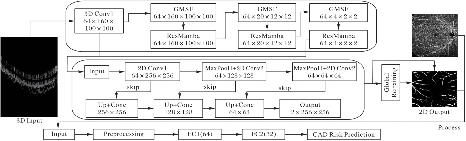
Fig. 1 Overall framework of MA_DNet
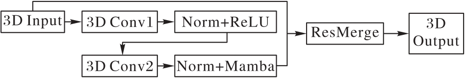
Fig. 2 ResMamba module structure
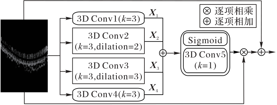
Fig. 3 GMSF module structure
| 子集 | MA_Net | MA_Net+ | |||||||
|---|---|---|---|---|---|---|---|---|---|
| 输入体积 | 分割块大小 | 批量大小 | 迭代次数 | 模型保存频率 | 输入图片大小 | 批量大小 | 迭代次数 | 模型保存频率 | |
| OCTA_3M | 304×304×640 | 76×76×160 | 3 | 25 000 | 300∶1 | 304×304 | 2 | 3 000 | 50∶1 |
| OCTA_6M | 400×600×640 | 100×100×160 | 3 | 25 000 | 300∶1 | 400×400 | 2 | 3 000 | 50∶1 |
Tab. 1 Network configuration of MA_Net and MA_Net+ on two subsets
| 子集 | MA_Net | MA_Net+ | |||||||
|---|---|---|---|---|---|---|---|---|---|
| 输入体积 | 分割块大小 | 批量大小 | 迭代次数 | 模型保存频率 | 输入图片大小 | 批量大小 | 迭代次数 | 模型保存频率 | |
| OCTA_3M | 304×304×640 | 76×76×160 | 3 | 25 000 | 300∶1 | 304×304 | 2 | 3 000 | 50∶1 |
| OCTA_6M | 400×600×640 | 100×100×160 | 3 | 25 000 | 300∶1 | 400×400 | 2 | 3 000 | 50∶1 |
| 基线 | GMSF | ResMamba | Dice | Jac | Bacc | Pre | Rec |
|---|---|---|---|---|---|---|---|
| ✓ | 91.71 | 84.80 | 95.36 | 92.29 | 89.43 | ||
| ✓ | ✓ | 90.55 | 82.88 | 94.72 | 91.27 | 90.07 | |
| ✓ | ✓ | 92.05 | 85.38 | 95.07 | 93.70 | 90.58 | |
| ✓ | ✓ | ✓ | 92.77 | 86.60 | 95.67 | 93.89 | 91.78 |
Tab. 2 Ablation experiment results on OCTA_3M subset
| 基线 | GMSF | ResMamba | Dice | Jac | Bacc | Pre | Rec |
|---|---|---|---|---|---|---|---|
| ✓ | 91.71 | 84.80 | 95.36 | 92.29 | 89.43 | ||
| ✓ | ✓ | 90.55 | 82.88 | 94.72 | 91.27 | 90.07 | |
| ✓ | ✓ | 92.05 | 85.38 | 95.07 | 93.70 | 90.58 | |
| ✓ | ✓ | ✓ | 92.77 | 86.60 | 95.67 | 93.89 | 91.78 |
| 基线 | GMSF | ResMamba | Dice | Jac | Bacc | Pre | Rec |
|---|---|---|---|---|---|---|---|
| ✓ | 88.19 | 78.98 | 93.22 | 88.99 | 87.57 | ||
| ✓ | ✓ | 88.82 | 80.02 | 92.09 | 93.46 | 84.80 | |
| ✓ | ✓ | 89.05 | 80.38 | 92.58 | 92.67 | 85.88 | |
| ✓ | ✓ | ✓ | 89.40 | 80.94 | 93.81 | 90.40 | 88.62 |
Tab. 3 Ablation experiment results on OCTA_6M subset
| 基线 | GMSF | ResMamba | Dice | Jac | Bacc | Pre | Rec |
|---|---|---|---|---|---|---|---|
| ✓ | 88.19 | 78.98 | 93.22 | 88.99 | 87.57 | ||
| ✓ | ✓ | 88.82 | 80.02 | 92.09 | 93.46 | 84.80 | |
| ✓ | ✓ | 89.05 | 80.38 | 92.58 | 92.67 | 85.88 | |
| ✓ | ✓ | ✓ | 89.40 | 80.94 | 93.81 | 90.40 | 88.62 |
| 模型 | Dice | Jac | Bacc |
|---|---|---|---|
| U-Net[ | 88.55±3.43 | 79.62±5.08 | 93.31±1.68 |
| U-Net++[ | 88.61±3.07 | 79.67±4.59 | 91.93±1.74 |
| Attention U-Net[ | 88.71±3.19 | 79.84±4.74 | 93.14±1.49 |
| Joint-seg[ | 91.13±2.09 | 83.78±3.40 | — |
| IPN[ | 90.62±5.96 | 83.25±7.78 | 93.87±4.19 |
| IPN-V2[ | 92.46±3.93 | 86.19±5.83 | 95.34±3.16 |
| IPN-V2+[ | 92.74±3.95 | 86.67±5.88 | 95.22±3.12 |
| FARGO[ | 91.68±2.05 | 84.70±3.34 | — |
| MA_Net | 92.77±2.43 | 86.60±3.90 | 95.67±2.07 |
| MA_Net+ | 93.02±2.40 | 87.04±3.89 | 95.80±2.00 |
Tab. 4 Comparison results of proposed and other OCTA models on OCTA_3M subset
| 模型 | Dice | Jac | Bacc |
|---|---|---|---|
| U-Net[ | 88.55±3.43 | 79.62±5.08 | 93.31±1.68 |
| U-Net++[ | 88.61±3.07 | 79.67±4.59 | 91.93±1.74 |
| Attention U-Net[ | 88.71±3.19 | 79.84±4.74 | 93.14±1.49 |
| Joint-seg[ | 91.13±2.09 | 83.78±3.40 | — |
| IPN[ | 90.62±5.96 | 83.25±7.78 | 93.87±4.19 |
| IPN-V2[ | 92.46±3.93 | 86.19±5.83 | 95.34±3.16 |
| IPN-V2+[ | 92.74±3.95 | 86.67±5.88 | 95.22±3.12 |
| FARGO[ | 91.68±2.05 | 84.70±3.34 | — |
| MA_Net | 92.77±2.43 | 86.60±3.90 | 95.67±2.07 |
| MA_Net+ | 93.02±2.40 | 87.04±3.89 | 95.80±2.00 |
| 模型 | Dice | Jac | Bacc |
|---|---|---|---|
| U-Net[ | 88.55±3.43 | 79.62±5.08 | 93.31±1.68 |
| U-Net++[ | 88.61±3.07 | 79.67±4.59 | 91.93±1.74 |
| Attention U-Net[ | 88.71±3.19 | 79.84±4.74 | 93.14±1.49 |
| Joint-seg[ | 89.72±3.21 | 81.17±3.11 | — |
| IPN[ | 88.64±3.21 | 79.73±4.92 | 93.07±2.42 |
| IPN-V2[ | 89.08±2.73 | 80.41±4.29 | 93.52±2.13 |
| IPN-V2+[ | 89.41±2.74 | 80.95±4.32 | 93.46±2.12 |
| PAENet[ | 89.36±2.70 | 80.43±4.15 | 94.05±1.95 |
| PAENet+[ | 89.69±2.77 | 81.42±4.39 | 93.68±2.08 |
| FARGO[ | 89.15±2.39 | 80.50±3.75 | — |
| MA_Net | 89.40±2.73 | 80.94±4.27 | 93.81±2.09 |
| MA_Net+ | 89.76±2.64 | 81.52±4.16 | 94.09±1.77 |
Tab. 5 Comparison results of proposed and other OCTA models on OCTA_6M subset
| 模型 | Dice | Jac | Bacc |
|---|---|---|---|
| U-Net[ | 88.55±3.43 | 79.62±5.08 | 93.31±1.68 |
| U-Net++[ | 88.61±3.07 | 79.67±4.59 | 91.93±1.74 |
| Attention U-Net[ | 88.71±3.19 | 79.84±4.74 | 93.14±1.49 |
| Joint-seg[ | 89.72±3.21 | 81.17±3.11 | — |
| IPN[ | 88.64±3.21 | 79.73±4.92 | 93.07±2.42 |
| IPN-V2[ | 89.08±2.73 | 80.41±4.29 | 93.52±2.13 |
| IPN-V2+[ | 89.41±2.74 | 80.95±4.32 | 93.46±2.12 |
| PAENet[ | 89.36±2.70 | 80.43±4.15 | 94.05±1.95 |
| PAENet+[ | 89.69±2.77 | 81.42±4.39 | 93.68±2.08 |
| FARGO[ | 89.15±2.39 | 80.50±3.75 | — |
| MA_Net | 89.40±2.73 | 80.94±4.27 | 93.81±2.09 |
| MA_Net+ | 89.76±2.64 | 81.52±4.16 | 94.09±1.77 |

Fig. 4 Visual comparison of segmentation results on OCTA_3M subset using different models
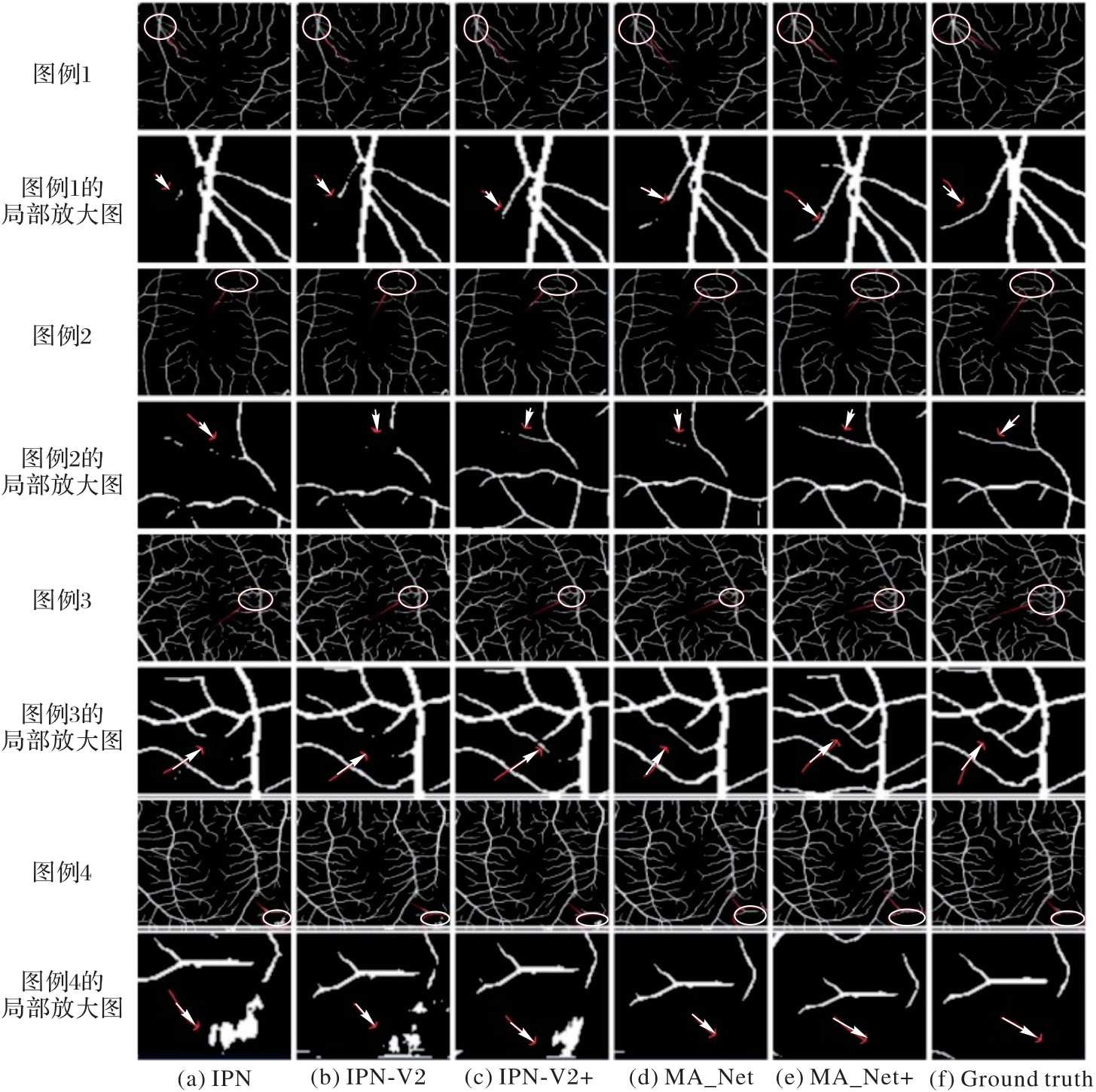
Fig. 5 Visual comparison of segmentation results on OCTA_6M subset using different models
| 模型 | 总参数量/106 | 模型 | 总参数量/106 |
|---|---|---|---|
| IPN-V2[ | 2.25 | IPN-V2+ResMamba | 2.33 |
| IPN-V2+[ | 3.81 | MA_Net | 3.36 |
| IPN-V2+GMSF | 2.34 | MA_Net+ | 4.86 |
Tab. 6 Model complexity comparison of existing networks on OCTA-500 dataset
| 模型 | 总参数量/106 | 模型 | 总参数量/106 |
|---|---|---|---|
| IPN-V2[ | 2.25 | IPN-V2+ResMamba | 2.33 |
| IPN-V2+[ | 3.81 | MA_Net | 3.36 |
| IPN-V2+GMSF | 2.34 | MA_Net+ | 4.86 |
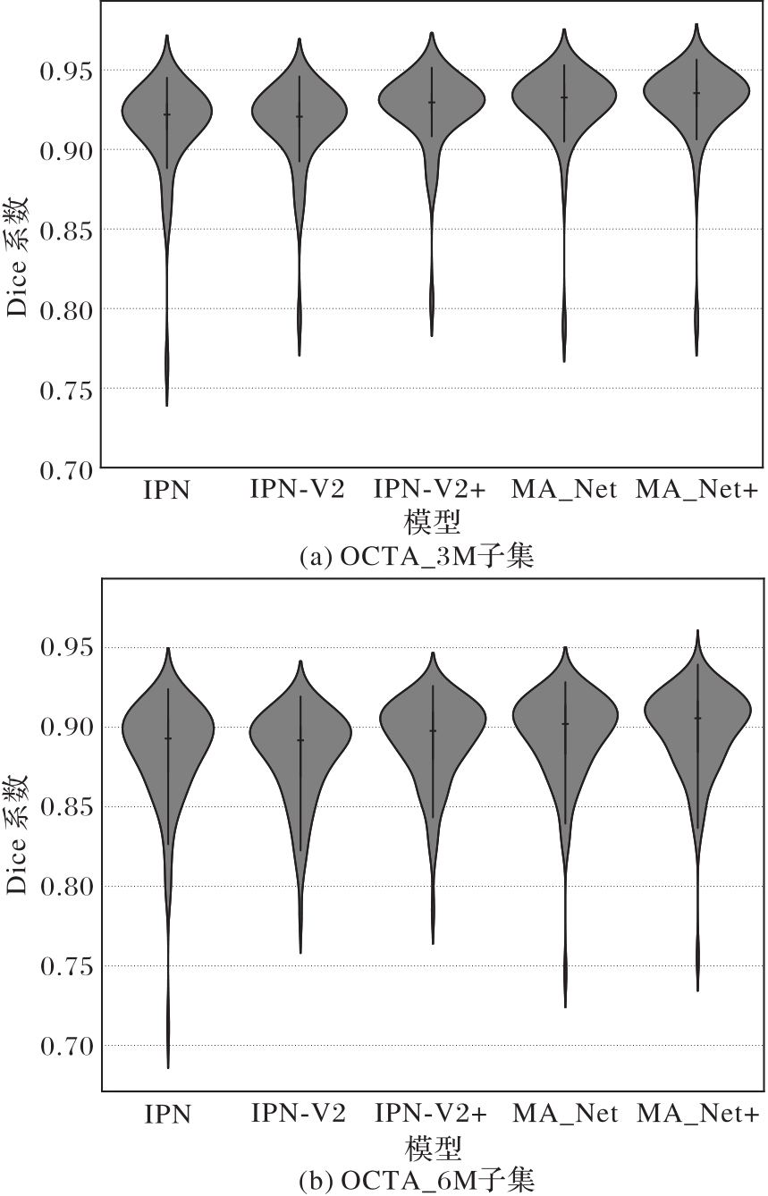
Fig. 6 Violin plots of Dice coefficient of various models
| 指标 | 数值 | 指标 | 数值 |
|---|---|---|---|
| 准确率 | 71.93 | Bacc | 71.71 |
| AUC | 86.23 | 召回率 | 69.23 |
Tab. 7 Prediction results of CAD
| 指标 | 数值 | 指标 | 数值 |
|---|---|---|---|
| 准确率 | 71.93 | Bacc | 71.71 |
| AUC | 86.23 | 召回率 | 69.23 |
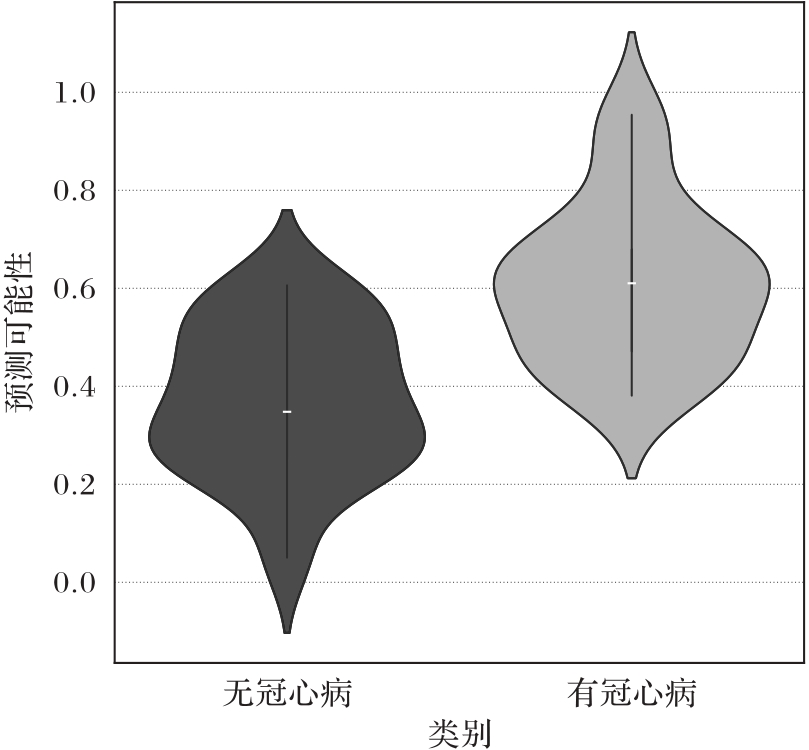
Fig. 7 Violin plot of coronary artery disease prediction
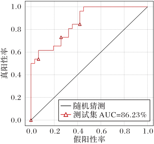
Fig. 8 ROC curves and AUC values for coronary artery disease
| [1] | WINTHER S, SCHMIDT S E, MAYRHOFER T, et al. Incorporating coronary calcification into pre-test assessment of the likelihood of coronary artery disease[J]. Journal of the American College of Cardiology, 2020, 76(21): 2421-2432. |
| [2] | MASSIN P, ERGINAY A, HAOUCHINE B, et al. Retinal thickness in healthy and diabetic subjects measured using optical coherence tomography mapping software[J]. European Journal of Ophthalmology, 2002, 12(2): 102-108. |
| [3] | LAVIA C, BONNIN S, MAULE M, et al. Vessel density of superficial, intermediate, and deep capillary plexuses using optical coherence tomography angiography[J]. Retina, 2019, 39(2): 247-258. |
| [4] | VILLAPLANA-VELASCO A, PIGEYRE M, ENGELMANN J, et al. Fine-mapping of retinal vascular complexity loci identifies notch regulation as a shared mechanism with myocardial infarction outcomes[J]. Communications Biology, 2023, 6: No.523. |
| [5] | WONG T Y, ISLAM F M A, KLEIN R, et al. Retinal vascular caliber, cardiovascular risk factors, and inflammation: the Multi-Ethnic Study of Atherosclerosis (MESA)[J]. Investigative Ophthalmology and Visual Science, 2006, 47(6): 2341-2350. |
| [6] | MacGILLIVRAY T J, TRUCCO E, CAMERON J R, et al. Retinal imaging as a source of biomarkers for diagnosis, characterization and prognosis of chronic illness or long-term conditions[J]. The British Journal of Radiology, 2014, 87(1040): No.20130832. |
| [7] | WANG S B, MITCHELL P, LIEW G, et al. A spectrum of retinal vasculature measures and coronary artery disease[J]. Atherosclerosis, 2018, 268: 215-224. |
| [8] | ISTVÁN L, CZAKÓ C, ÉLŐ Á, et al. Imaging retinal microvascular manifestations of carotid artery disease in older adults: from diagnosis of ocular complications to understanding microvascular contributions to cognitive impairment[J]. GeroScience, 2021, 43(4): 1703-1723. |
| [9] | ARNOULD L, GUENANCIA C, BINQUET C, et al. Caractéristiques vasculaires rétiniennes: modifications lors du vieillissement et en pathologie vasculaire systémique (cardiaque et cérébrale)[J]. Journal Français d’Ophtalmologie, 2022, 45(1): 104-118. |
| [10] | IORGA R E, COSTIN D, MUNTEANU-DĂNULESCU R S, et al. Non-invasive retinal vessel analysis as a predictor for cardiovascular disease[J]. Journal of Personalized Medicine, 2024, 14(5): No.501. |
| [11] | LEE S J V, GOH Y Q, ROJAS-CARABALI W, et al. Association between retinal vessels caliber and systemic health: a comprehensive review[J]. Survey of Ophthalmology, 2025, 70(2): 184-199. |
| [12] | ARNOULD L, MERIAUDEAU F, GUENANCIA C, et al. Using artificial intelligence to analyse the retinal vascular network: the future of cardiovascular risk assessment based on oculomics? a narrative review[J]. Ophthalmology and Therapy, 2023, 12(2): 657-674. |
| [13] | SHIROMANI S, AlBADRI A, LINDEKE-MYERS A, et al. Reduced retinal microvascular density in women with coronary microvascular dysfunction: a pilot study[J]. American Heart Journal Plus: Cardiology Research and Practice, 2025, 51: No.100502. |
| [14] | ZEPPENFELD K, TFELT-HANSEN J, DE RIVA M, et al. 2022 ESC Guidelines for the management of patients with ventricular arrhythmias and the prevention of sudden cardiac death: developed by the task force for the management of patients with ventricular arrhythmias and the prevention of sudden cardiac death of the European Society of Cardiology (ESC) Endorsed by the Association for European Paediatric and Congenital Cardiology (AEPC)[J]. European Heart Journal, 2022, 43(40): 3997-4126. |
| [15] | HU D, PAN L, CHEN X, et al. A novel vessel segmentation algorithm for pathological en-face images based on matched filter[J]. Physics in Medicine and Biology, 2023, 68(5): No.055014. |
| [16] | GHENCIU L A, DIMA M, STOICESCU E R, et al. Retinal imaging-based oculomics: artificial intelligence as a tool in the diagnosis of cardiovascular and metabolic diseases[J]. Biomedicines, 2024, 12(9): No.2150. |
| [17] | LISA GRACIA M, VIEITEZ SANTIAGO M, SALMÓN GONZALEZ Z, et al. La hipertensión arterial y el score de Framingham de riesgo vascular en la obstrucción venosa retiniana[J]. Hipertensión y Riesgo Vascular, 2019, 36(4): 193-198. |
| [18] | VASWANI A, SHAZEER N, PARMAR N, et al. Attention is all you need[C]// Proceedings of the 31st International Conference on Neural Information Processing Systems. Red Hook: Curran Associates Inc., 2017: 6000-6010. |
| [19] | DEVLIN J, CHANG M W, LEE K, et al. BERT: pre-training of deep bidirectional Transformers for language understanding[C]// Proceedings of the 2019 Conference of the North American Chapter of the Association for Computational Linguistics: Human Language Technologies, Volume 1 (Long and Short Papers). Stroudsburg: ACL, 2019: 4171-4186. |
| [20] | XING Z, YU L, WAN L, et al. NestedFormer: nested modality-aware Transformer for brain tumor segmentation[EB/OL]. [2024-11-05].. |
| [21] | GU A, DAO T. Mamba: linear-time sequence modeling with selective state spaces[EB/OL]. [2024-10-12].. |
| [22] | LIU Y, TIAN Y, ZHAO Y, et al. VMamba: visual state space model[EB/OL]. [2024-09-30].. |
| [23] | BASAR T. A new approach to linear filtering and prediction problems[M]. [S.l.]:Wiley-IEEE Press, 2001: 167-179. |
| [24] | MA J, LI F, WANG B. U-Mamba: enhancing long-range dependency for biomedical image segmentation[EB/OL]. [2024-12-15].. |
| [25] | 孙颖,丁卫平,黄嘉爽,等. RCAR-UNet:基于粗糙通道注意力机制的视网膜血管分割网络[J]. 计算机研究与发展, 2023, 60(4): 947-961. |
| SUN Y, DING W P, HUANG J S, et al. RCAR-UNet: retinal vessels segmentation network based on rough channel attention mechanism[J]. Journal of Computer Research and Development, 2023, 60(4): 947-961. | |
| [26] | 梁礼明,黄朝林,石霏,等. 融合形状先验的水平集眼底图像血管分割[J]. 计算机学报, 2018, 41(7): 1678-1692. |
| LIANG L M, HUANG C L, SHI F, et al. Retinal vessel segmentation in fundus images using level set combined with shape priori[J]. Chinese Journal of Computers, 2018, 41(7): 1678-1692. | |
| [27] | 解立志,周明全,田沄,等. 基于区域增长与局部自适应C-V模型的脑血管分割[J]. 软件学报, 2013, 24(8): 1927-1936. |
| XIE L Z, ZHOU M Q, TIAN Y, et al. Cerebrovascular segmentation based on region growing and local adaptive C-V model[J]. Journal of Software, 2013, 24(8): 1927-1936. | |
| [28] | 梁礼明,刘博文,杨海龙,等. 基于多特征融合的有监督视网膜血管提取[J]. 计算机学报, 2018, 41(11): 2566-2580. |
| LIANG L M, LIU B W, YANG H L, et al. Supervised blood vessel extraction in retinal images based on multi-feature fusion[J]. Chinese Journal of Computers, 2018, 41(11): 2566-2580. | |
| [29] | MAIER H, FAGHIHROOHI S, NAVAB N. A line to align: deep dynamic time warping for retinal OCT segmentation[C]// Proceedings of the 2021 International Conference on Medical Image Computing and Computer-Assisted Intervention, LNCS 12901. Cham: Springer, 2021: 709-719. |
| [30] | LI M, CHEN Y, JI Z, et al. Image projection network: 3D to 2D image segmentation in OCTA images[J]. IEEE Transactions on Medical Imaging, 2020, 39(11): 3343-3354. |
| [31] | LI M, HUANG K, XU Q, et al. OCTA-500: a retinal dataset for optical coherence tomography angiography study[J]. Medical Image Analysis, 2024, 93: No.103092. |
| [32] | QUAN X, HOU G, YIN W, et al. A multi-modal and multi-stage fusion enhancement network for segmentation based on OCT and OCTA images[J]. Information Fusion, 2025, 113: No.102594. |
| [33] | YANG C, LI B, XIAO Q, et al. LA-Net: layer attention network for 3D-to-2D retinal vessel segmentation in OCTA images[J]. Physics in Medicine and Biology, 2024, 69(4): No.045019. |
| [34] | TUN Y Z, AIMMANEE P. Machine learning-based retinal neovascularization localization in en face optical coherence tomography angiography images using vessel features[J]. IEEE Access, 2025, 13: 2045-2057. |
| [35] | FRAZ M M, REMAGNINO P, HOPPE A, et al. Blood vessel segmentation methodologies in retinal images: a survey[J]. Computer Methods and Programs in Biomedicine, 2012, 108(1): 407-433. |
| [36] | BRUMMER A B, HUNT D, SAVAGE V. Improving blood vessel tortuosity measurements via highly sampled numerical integration of the Frenet-Serret equations[J]. IEEE Transactions on Medical Imaging, 2021, 40(1): 297-309. |
| [37] | CHE AZEMIN M Z, HAMID F AB, WANG J J, et al. Box-counting fractal dimension algorithm variations on retina images[C]// Advanced Computer and Communication Engineering Technology: Proceedings of ICOCOE 2015, LNEE 362. Cham: Springer, 2016: 337-343. |
| [38] | DASH J, BHOI N. Retinal blood vessel segmentation using Otsu thresholding with principal component analysis[C]// Proceedings of the 2nd International Conference on Inventive Systems and Control. Piscataway: IEEE, 2018: 933-937. |
| [39] | NAGASATO D, TABUCHI H, MASUMOTO H, et al. Automated detection of a nonperfusion area caused by retinal vein occlusion in optical coherence tomography angiography images using deep learning[J]. PLoS ONE, 2019, 14(11): No.e0223965. |
| [40] | LUISI J, LIU W, ZHANG W, et al. En-face optical coherence tomography angiography for longitudinal monitoring of retinal injury[J]. Applied Sciences, 2019, 9(13): No.2617. |
| [41] | HUANG H, LIN L, TONG R, et al. UNet 3+: a full-scale connected UNet for medical image segmentation[C]// Proceedings of the 2020 IEEE International Conference on Acoustics, Speech and Signal Processing. Piscataway: IEEE, 2020: 1055-1059. |
| [42] | RONNEBERGER O, FISCHER P, BROX T. U-Net: convolutional networks for biomedical image segmentation[C]// Proceedings of the 2015 International Conference on Medical Image Computing and Computer-Assisted Intervention, LNCS 9351. Cham: Springer, 2015: 234-241. |
| [43] | ZHOU Z, RAHMAN SIDDIQUEE M M, TAJBAKHSH N, et al. UNet++: a nested U-Net architecture for medical image segmentation[C]// Proceedings of the 2018 International Workshop on Deep Learning in Medical Image Analysis, LNCS 11045. Cham: Springer, 2018: 3-11. |
| [44] | OKTAY O, SCHLEMPER J, FOLGOC L L, et al. Attention U-Net: learning where to look for the pancreas[EB/OL]. [2024-11-28].. |
| [45] | HU K, JIANG S, ZHANG Y, et al. Joint-Seg: treat foveal avascular zone and retinal vessel segmentation in OCTA images as a joint task[J]. IEEE Transactions on Instrumentation and Measurement, 2022, 71: No.4007113. |
| [46] | WU Z, WANG Z, ZOU W, et al. PAENet: a progressive attention-enhanced network for 3D to 2D retinal vessel segmentation[C]// Proceedings of the 2021 IEEE International Conference on Bioinformatics and Biomedicine. Piscataway: IEEE, 2021: 1579-1584. |
| [47] | PENG L, LIN L, CHENG P, et al. FARGO: a joint framework for FAZ and RV segmentation from OCTA images[C]// Proceedings of the 2021 International Workshop on Ophthalmic Medical Image Analysis. Cham: Springer, 2021: 42-51. |
| [48] | LIU M, LOVERN C, LYCETT K, et al. The association between markers of inflammation and retinal microvascular parameters: a systematic review and meta-analysis[J]. Atherosclerosis, 2021, 336: 12-22. |
| [1] | Haoqian JIANG, Dong ZHANG, Guanyu LI, Heng CHEN. SetaCRS: Conversational recommender system with structure-enhanced hierarchical task-oriented prompting strategy [J]. Journal of Computer Applications, 2026, 46(2): 368-377. |
| [2] | Feng HAN, Yongfeng BU, Haoxiang LIANG, Shuwen HUANG, Zhaoyang ZHANG, Shijie SUN. Vehicle trajectory anomaly detection based on multi-level spatio-temporal interaction dependency [J]. Journal of Computer Applications, 2026, 46(2): 604-612. |
| [3] | Xiaolei CHEN, Zhiwei ZHENG, Xue HUANG, Zhenbin QU. Panoramic video super-resolution network combining spherical alignment and adaptive geometric correction [J]. Journal of Computer Applications, 2026, 46(2): 528-535. |
| [4] | Quanjie LIU, Zhaoyi GU, Chunyuan WANG. Unsafe driving behavior detection under complex lighting conditions [J]. Journal of Computer Applications, 2026, 46(2): 613-619. |
| [5] | Jinjiao LIN, Canshun ZHANG, Shuya CHEN, Tianxin WANG, Jian LIAN, Yonghui XU. Vehicle insurance fraud detection method based on improved graph attention network [J]. Journal of Computer Applications, 2026, 46(2): 437-444. |
| [6] | Ming LI, Mengqi WANG, Aili ZHANG, Hua REN, Yuqiang DOU. Image steganography method based on conditional generative adversarial networks and hybrid attention mechanism [J]. Journal of Computer Applications, 2026, 46(2): 475-484. |
| [7] | Jie CAO, Lingfeng XIE, Bingjin WANG, Changhe ZHANG, Zidong YU, Chao DENG. Adolescent scoliosis screening method considering class imbalance and background diversity [J]. Journal of Computer Applications, 2026, 46(2): 630-639. |
| [8] | Zeyi GUO, Fenglian LI, Lichun XU. Double decision mechanism-based deep symbolic regression algorithm [J]. Journal of Computer Applications, 2026, 46(2): 406-415. |
| [9] | Xiaoyong BIAN, Peiyang YUAN, Qiren HU. Dual-coding space-frequency mixing method for infrared small target detection [J]. Journal of Computer Applications, 2026, 46(1): 252-259. |
| [10] | Zhihui ZAN, Yajing WANG, Ke LI, Zhixiang YANG, Guangyu YANG. Multi-feature fusion speech emotion recognition method based on SAA-CNN-BiLSTM network [J]. Journal of Computer Applications, 2026, 46(1): 69-76. |
| [11] | Panfeng JING, Yudong LIANG, Chaowei LI, Junru GUO, Jinyu GUO. Semi-supervised image dehazing algorithm based on teacher-student learning [J]. Journal of Computer Applications, 2025, 45(9): 2975-2983. |
| [12] | Hongjun ZHANG, Gaojun PAN, Hao YE, Yubin LU, Yiheng MIAO. Multi-source heterogeneous data analysis method combining deep learning and tensor decomposition [J]. Journal of Computer Applications, 2025, 45(9): 2838-2847. |
| [13] | Jin LI, Liqun LIU. SAR and visible image fusion based on residual Swin Transformer [J]. Journal of Computer Applications, 2025, 45(9): 2949-2956. |
| [14] | Bing YIN, Zhenhua LING, Yin LIN, Changfeng XI, Ying LIU. Emotion recognition method compatible with missing modal reasoning [J]. Journal of Computer Applications, 2025, 45(9): 2764-2772. |
| [15] | Weigang LI, Jiale SHAO, Zhiqiang TIAN. Point cloud classification and segmentation network based on dual attention mechanism and multi-scale fusion [J]. Journal of Computer Applications, 2025, 45(9): 3003-3010. |
| Viewed | ||||||
|
Full text |
|
|||||
|
Abstract |
|
|||||