


Journal of Computer Applications ›› 2026, Vol. 46 ›› Issue (1): 280-288.DOI: 10.11772/j.issn.1001-9081.2024121868
• Multimedia computing and computer simulation • Previous Articles Next Articles
Chaoyun MAI1, Hongyi ZHANG1, Chuanbo QIN1( ), Junying ZENG1, Dong WANG2
Received:2025-01-03
Revised:2025-03-21
Accepted:2025-03-25
Online:2026-01-10
Published:2026-01-10
Contact:
Chuanbo QIN
About author:MAI Chaoyun, born in 1989, Ph. D., associate professor. His research interests include intelligent information processing, digital signal processing.Supported by:
麦超云1, 张洪燚1, 秦传波1(), 曾军英1, 王栋2
通讯作者:
秦传波
作者简介:麦超云(1989—),男,广东江门人,副教授,博士,CCF会员,主要研究方向:智能信息处理、数字信号处理基金资助:CLC Number:
Chaoyun MAI, Hongyi ZHANG, Chuanbo QIN, Junying ZENG, Dong WANG. Multi-scale and spatial frequency feature-based image segmentation network for pheochromocytoma[J]. Journal of Computer Applications, 2026, 46(1): 280-288.
麦超云, 张洪燚, 秦传波, 曾军英, 王栋. 基于多尺度与空间频率特征的嗜铬细胞瘤图像分割网络[J]. 《计算机应用》唯一官方网站, 2026, 46(1): 280-288.
Add to citation manager EndNote|Ris|BibTeX
URL: https://www.joca.cn/EN/10.11772/j.issn.1001-9081.2024121868
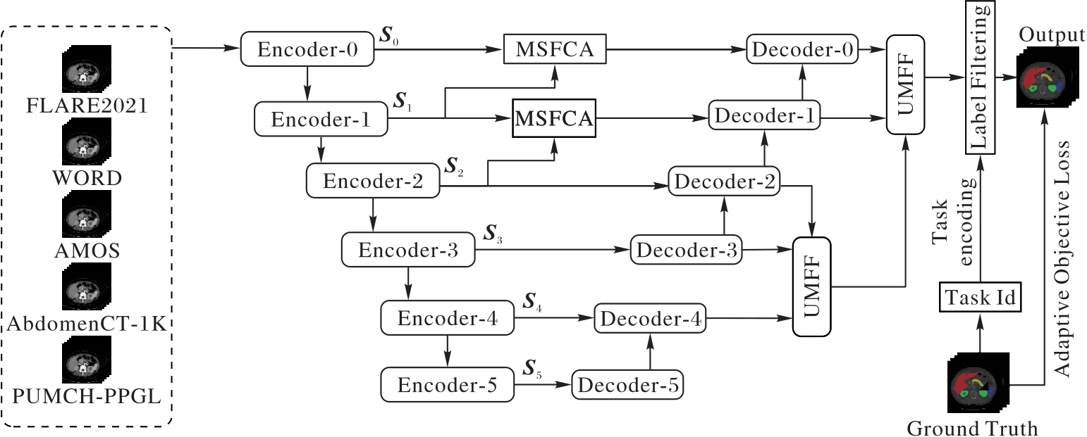
Fig. 1 Overall architecture of MF-Net
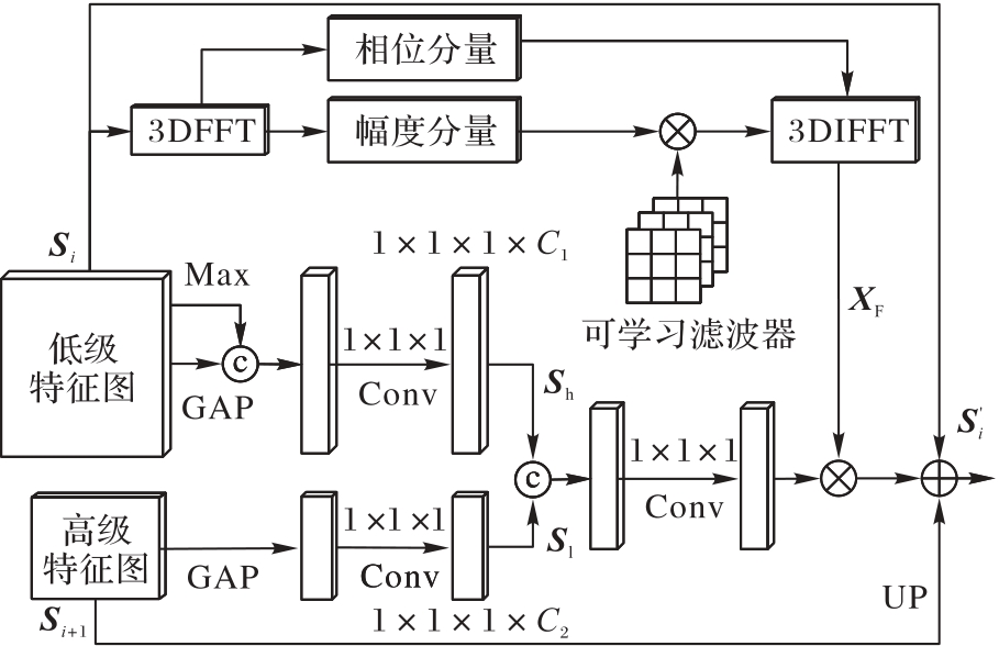
Fig. 2 Structure of MSFCA
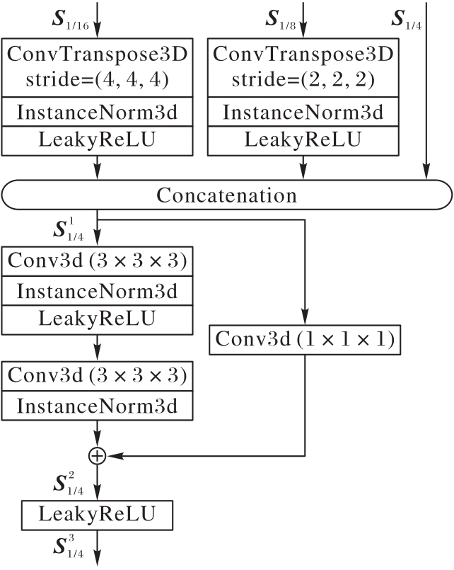
Fig. 3 Structure of UMFF
| 数据集 | 训练集样本数 | 测试集样本数 | 注释类别数 |
|---|---|---|---|
| AbdomenCT-1K[ | 800 | 200 | 4 |
| AMOS-CT[ | 240 | 60 | 13 |
| FLARE2021[ | 361 | 30 | 4 |
| WORD[ | 120 | 30 | 11 |
| BTCV[ | 0 | 30 | 12 |
| FLARE2022[ | 0 | 50 | 13 |
| PUMCH-PPGL | 150 | 42 | 1 |
Tab. 1 Abdominal organ datasets and annotated organs
| 数据集 | 训练集样本数 | 测试集样本数 | 注释类别数 |
|---|---|---|---|
| AbdomenCT-1K[ | 800 | 200 | 4 |
| AMOS-CT[ | 240 | 60 | 13 |
| FLARE2021[ | 361 | 30 | 4 |
| WORD[ | 120 | 30 | 11 |
| BTCV[ | 0 | 30 | 12 |
| FLARE2022[ | 0 | 50 | 13 |
| PUMCH-PPGL | 150 | 42 | 1 |
| 评价指标 | 方法 | 肝脏 | 右肾 | 脾 | 胰腺 | 主动脉 | 下腔 静脉 | 右肾 上腺 | 左肾 上腺 | 胆囊 | 食管 | 胃 | 十二 指肠 | 左肾 | PPGL | 平均 |
|---|---|---|---|---|---|---|---|---|---|---|---|---|---|---|---|---|
| Dice | Multi-Net[ | 95.14 | 90.86 | 92.29 | 78.78 | 87.83 | 91.52 | 78.11 | 83.33 | 82.70 | 82.41 | 81.55 | 75.37 | 90.34 | 90.08 | 85.74 |
| TAL[ | 94.47 | 92.39 | 93.04 | 82.03 | 90.33 | 91.03 | 78.28 | 79.69 | 83.59 | 82.82 | 84.84 | 82.08 | 91.71 | 86.57 | 86.63 | |
| Multi-head[ | 94.94 | 89.46 | 94.97 | 84.18 | 85.62 | 94.02 | 80.32 | 82.65 | 86.82 | 84.09 | 74.48 | 78.90 | 89.67 | 88.43 | 86.33 | |
| CLIP-Driven[ | 96.57 | 91.72 | 94.66 | 84.33 | 89.47 | 92.41 | 77.84 | 79.92 | 86.01 | 84.70 | 89.25 | 80.80 | 93.13 | 90.14 | 87.93 | |
| DoDNet[ | 96.80 | 92.15 | 94.54 | 86.13 | 90.12 | 92.66 | 77.05 | 80.10 | 83.57 | 84.06 | 90.39 | 82.77 | 92.26 | 90.02 | 88.04 | |
| MF-Net | 96.92 | 93.31 | 94.56 | 86.41 | 89.83 | 93.78 | 80.42 | 83.49 | 87.37 | 84.18 | 89.63 | 82.15 | 94.65 | 90.31 | 89.07 | |
| NSD | Multi-Net[ | 95.32 | 92.41 | 93.44 | 87.21 | 94.69 | 90.27 | 84.39 | 87.71 | 86.20 | 83.93 | 90.27 | 88.56 | 88.86 | 92.08 | 89.67 |
| TAL[ | 93.33 | 93.67 | 92.79 | 89.80 | 96.85 | 94.21 | 85.73 | 86.08 | 84.11 | 87.45 | 91.62 | 86.37 | 93.67 | 90.26 | 90.42 | |
| Multi-head[ | 95.12 | 92.46 | 94.68 | 88.08 | 97.83 | 94.15 | 87.36 | 86.83 | 85.45 | 84.08 | 88.68 | 89.61 | 86.08 | 88.37 | 89.91 | |
| CLIP-Driven[ | 97.44 | 93.15 | 95.83 | 89.62 | 96.46 | 93.83 | 86.11 | 85.24 | 87.74 | 91.37 | 92.52 | 92.32 | 90.64 | 92.14 | 91.74 | |
| DoDNet[ | 97.75 | 93.56 | 95.77 | 91.47 | 95.37 | 93.68 | 85.67 | 84.72 | 89.92 | 85.85 | 93.22 | 90.59 | 93.32 | 91.02 | 91.57 | |
| MF-Net | 97.79 | 94.36 | 96.28 | 91.86 | 97.36 | 94.32 | 87.54 | 88.21 | 91.42 | 91.13 | 93.62 | 91.52 | 92.15 | 92.40 | 92.85 |
Tab. 2 Segmentation results on retained test sets
| 评价指标 | 方法 | 肝脏 | 右肾 | 脾 | 胰腺 | 主动脉 | 下腔 静脉 | 右肾 上腺 | 左肾 上腺 | 胆囊 | 食管 | 胃 | 十二 指肠 | 左肾 | PPGL | 平均 |
|---|---|---|---|---|---|---|---|---|---|---|---|---|---|---|---|---|
| Dice | Multi-Net[ | 95.14 | 90.86 | 92.29 | 78.78 | 87.83 | 91.52 | 78.11 | 83.33 | 82.70 | 82.41 | 81.55 | 75.37 | 90.34 | 90.08 | 85.74 |
| TAL[ | 94.47 | 92.39 | 93.04 | 82.03 | 90.33 | 91.03 | 78.28 | 79.69 | 83.59 | 82.82 | 84.84 | 82.08 | 91.71 | 86.57 | 86.63 | |
| Multi-head[ | 94.94 | 89.46 | 94.97 | 84.18 | 85.62 | 94.02 | 80.32 | 82.65 | 86.82 | 84.09 | 74.48 | 78.90 | 89.67 | 88.43 | 86.33 | |
| CLIP-Driven[ | 96.57 | 91.72 | 94.66 | 84.33 | 89.47 | 92.41 | 77.84 | 79.92 | 86.01 | 84.70 | 89.25 | 80.80 | 93.13 | 90.14 | 87.93 | |
| DoDNet[ | 96.80 | 92.15 | 94.54 | 86.13 | 90.12 | 92.66 | 77.05 | 80.10 | 83.57 | 84.06 | 90.39 | 82.77 | 92.26 | 90.02 | 88.04 | |
| MF-Net | 96.92 | 93.31 | 94.56 | 86.41 | 89.83 | 93.78 | 80.42 | 83.49 | 87.37 | 84.18 | 89.63 | 82.15 | 94.65 | 90.31 | 89.07 | |
| NSD | Multi-Net[ | 95.32 | 92.41 | 93.44 | 87.21 | 94.69 | 90.27 | 84.39 | 87.71 | 86.20 | 83.93 | 90.27 | 88.56 | 88.86 | 92.08 | 89.67 |
| TAL[ | 93.33 | 93.67 | 92.79 | 89.80 | 96.85 | 94.21 | 85.73 | 86.08 | 84.11 | 87.45 | 91.62 | 86.37 | 93.67 | 90.26 | 90.42 | |
| Multi-head[ | 95.12 | 92.46 | 94.68 | 88.08 | 97.83 | 94.15 | 87.36 | 86.83 | 85.45 | 84.08 | 88.68 | 89.61 | 86.08 | 88.37 | 89.91 | |
| CLIP-Driven[ | 97.44 | 93.15 | 95.83 | 89.62 | 96.46 | 93.83 | 86.11 | 85.24 | 87.74 | 91.37 | 92.52 | 92.32 | 90.64 | 92.14 | 91.74 | |
| DoDNet[ | 97.75 | 93.56 | 95.77 | 91.47 | 95.37 | 93.68 | 85.67 | 84.72 | 89.92 | 85.85 | 93.22 | 90.59 | 93.32 | 91.02 | 91.57 | |
| MF-Net | 97.79 | 94.36 | 96.28 | 91.86 | 97.36 | 94.32 | 87.54 | 88.21 | 91.42 | 91.13 | 93.62 | 91.52 | 92.15 | 92.40 | 92.85 |
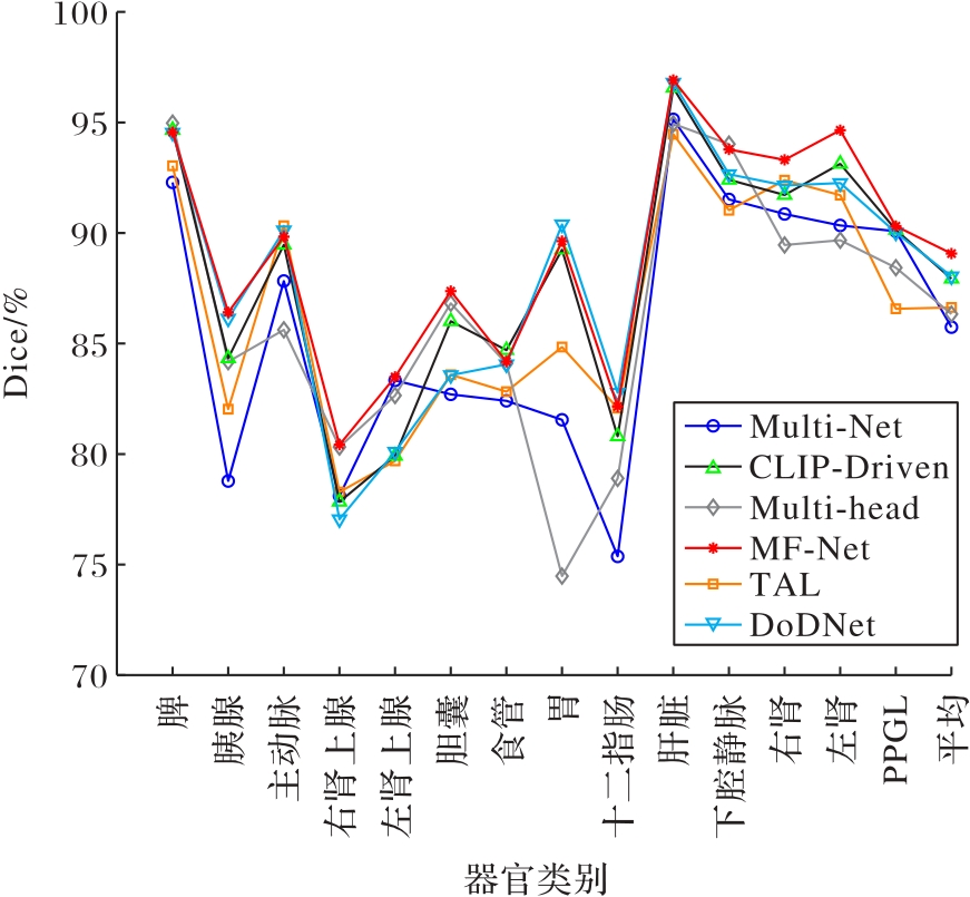
Fig. 4 Comparison of Dice on retained test sets

Fig. 5 Visualized segmentation results on retained test set
| 评价指标 | 方法 | 肝脏 | 右肾 | 脾 | 胰腺 | 主动脉 | 下腔 静脉 | 右肾 上腺 | 左肾 上腺 | 胆囊 | 食管 | 胃 | 十二 指肠 | 左肾 | 平均 |
|---|---|---|---|---|---|---|---|---|---|---|---|---|---|---|---|
| Dice | Multi-Net[ | 94.25 | 87.24 | 83.38 | 74.66 | 85.81 | 83.63 | 75.94 | 77.72 | 79.87 | 78.40 | 72.47 | 70.01 | 87.21 | 80.81 |
| TAL[ | 95.66 | 86.62 | 86.64 | 77.58 | 83.91 | 83.73 | 73.93 | 75.99 | 79.52 | 78.14 | 81.50 | 78.27 | 86.99 | 82.19 | |
| Multi-head[10] | 95.60 | 86.40 | 88.65 | 79.35 | 88.21 | 82.26 | 79.77 | 75.62 | 81.97 | 78.04 | 73.12 | 73.91 | 86.85 | 82.29 | |
| CLIP-Driven[ | 95.25 | 86.58 | 89.73 | 80.34 | 86.37 | 84.18 | 74.48 | 76.10 | 82.15 | 81.00 | 78.90 | 76.10 | 88.98 | 83.09 | |
| DoDNet[ | 95.38 | 87.61 | 90.59 | 79.56 | 87.27 | 84.76 | 76.23 | 76.72 | 81.23 | 79.67 | 80.73 | 76.90 | 87.94 | 83.43 | |
| MF-Net | 95.88 | 88.73 | 90.21 | 79.33 | 88.31 | 85.88 | 79.84 | 78.61 | 83.36 | 82.47 | 79.62 | 78.83 | 89.52 | 84.66 | |
| NSD | Multi-Net[ | 95.28 | 91.08 | 91.28 | 79.37 | 87.85 | 89.89 | 81.47 | 82.12 | 85.24 | 85.50 | 83.87 | 84.97 | 89.24 | 86.70 |
| TAL[ | 94.25 | 92.50 | 91.34 | 82.49 | 90.91 | 91.10 | 82.94 | 81.78 | 85.09 | 86.97 | 86.83 | 90.19 | 90.73 | 88.24 | |
| Multi-head[ | 95.34 | 89.99 | 93.54 | 83.33 | 88.15 | 92.90 | 84.92 | 84.32 | 87.83 | 86.59 | 78.59 | 86.04 | 88.51 | 87.70 | |
| CLIP-Driven[ | 97.05 | 91.80 | 93.60 | 83.88 | 90.43 | 92.17 | 82.57 | 81.46 | 87.89 | 89.99 | 90.28 | 89.28 | 92.28 | 89.44 | |
| DoDNet[ | 97.25 | 92.44 | 93.71 | 85.71 | 90.27 | 92.21 | 82.03 | 81.54 | 87.33 | 87.20 | 91.19 | 91.40 | 91.33 | 89.51 | |
| MF-Net | 97.34 | 93.28 | 93.38 | 86.07 | 91.36 | 92.30 | 84.97 | 84.46 | 88.15 | 89.81 | 90.77 | 91.21 | 93.99 | 90.55 |
Tab. 3 Test results on external datasets
| 评价指标 | 方法 | 肝脏 | 右肾 | 脾 | 胰腺 | 主动脉 | 下腔 静脉 | 右肾 上腺 | 左肾 上腺 | 胆囊 | 食管 | 胃 | 十二 指肠 | 左肾 | 平均 |
|---|---|---|---|---|---|---|---|---|---|---|---|---|---|---|---|
| Dice | Multi-Net[ | 94.25 | 87.24 | 83.38 | 74.66 | 85.81 | 83.63 | 75.94 | 77.72 | 79.87 | 78.40 | 72.47 | 70.01 | 87.21 | 80.81 |
| TAL[ | 95.66 | 86.62 | 86.64 | 77.58 | 83.91 | 83.73 | 73.93 | 75.99 | 79.52 | 78.14 | 81.50 | 78.27 | 86.99 | 82.19 | |
| Multi-head[10] | 95.60 | 86.40 | 88.65 | 79.35 | 88.21 | 82.26 | 79.77 | 75.62 | 81.97 | 78.04 | 73.12 | 73.91 | 86.85 | 82.29 | |
| CLIP-Driven[ | 95.25 | 86.58 | 89.73 | 80.34 | 86.37 | 84.18 | 74.48 | 76.10 | 82.15 | 81.00 | 78.90 | 76.10 | 88.98 | 83.09 | |
| DoDNet[ | 95.38 | 87.61 | 90.59 | 79.56 | 87.27 | 84.76 | 76.23 | 76.72 | 81.23 | 79.67 | 80.73 | 76.90 | 87.94 | 83.43 | |
| MF-Net | 95.88 | 88.73 | 90.21 | 79.33 | 88.31 | 85.88 | 79.84 | 78.61 | 83.36 | 82.47 | 79.62 | 78.83 | 89.52 | 84.66 | |
| NSD | Multi-Net[ | 95.28 | 91.08 | 91.28 | 79.37 | 87.85 | 89.89 | 81.47 | 82.12 | 85.24 | 85.50 | 83.87 | 84.97 | 89.24 | 86.70 |
| TAL[ | 94.25 | 92.50 | 91.34 | 82.49 | 90.91 | 91.10 | 82.94 | 81.78 | 85.09 | 86.97 | 86.83 | 90.19 | 90.73 | 88.24 | |
| Multi-head[ | 95.34 | 89.99 | 93.54 | 83.33 | 88.15 | 92.90 | 84.92 | 84.32 | 87.83 | 86.59 | 78.59 | 86.04 | 88.51 | 87.70 | |
| CLIP-Driven[ | 97.05 | 91.80 | 93.60 | 83.88 | 90.43 | 92.17 | 82.57 | 81.46 | 87.89 | 89.99 | 90.28 | 89.28 | 92.28 | 89.44 | |
| DoDNet[ | 97.25 | 92.44 | 93.71 | 85.71 | 90.27 | 92.21 | 82.03 | 81.54 | 87.33 | 87.20 | 91.19 | 91.40 | 91.33 | 89.51 | |
| MF-Net | 97.34 | 93.28 | 93.38 | 86.07 | 91.36 | 92.30 | 84.97 | 84.46 | 88.15 | 89.81 | 90.77 | 91.21 | 93.99 | 90.55 |
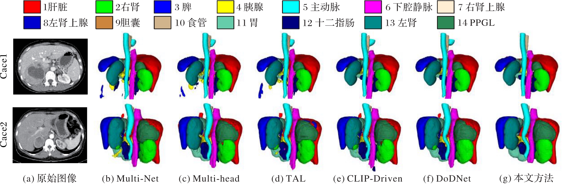
Fig. 6 Visualized segmentation results of pheochromocytoma images
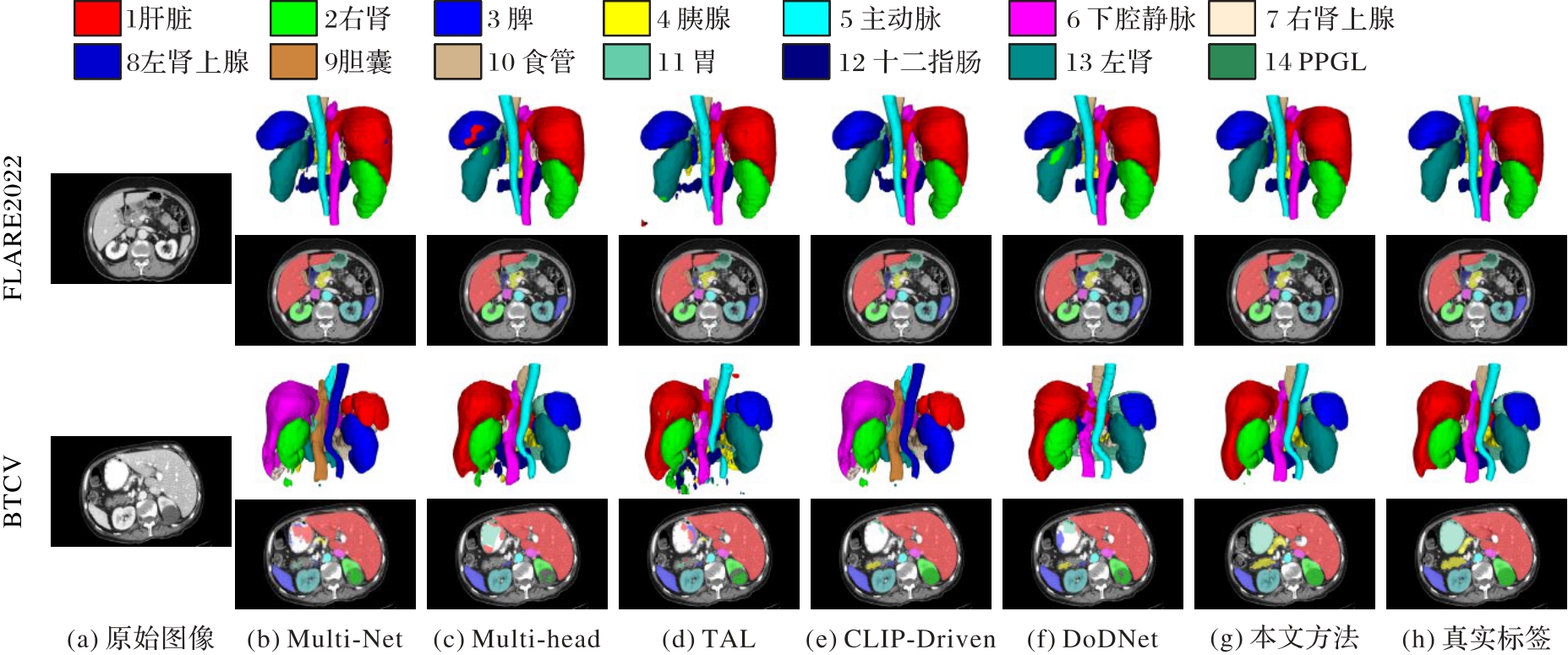
Fig. 7 Visualized segmentation results on BTCV and FLARE2022 datasets
| MSFCA | UMFF | AOb | AbdomenCT-1K | AMOS-CT | FLARE2021 | WORD | PUMCH-PPGL | 加权平均 | ||||||
|---|---|---|---|---|---|---|---|---|---|---|---|---|---|---|
| Dice | NSD | Dice | NSD | Dice | NSD | Dice | NSD | Dice | NSD | Dice | NSD | |||
| 90.02 | 93.33 | 32.85 | 34.69 | 80.97 | 83.64 | 41.25 | 43.95 | 65.31 | 67.89 | |||||
| √ | 90.36 | 93.78 | 88.27 | 91.95 | 81.36 | 83.95 | 81.64 | 86.89 | 88.65 | 92.78 | 88.35 | 91.98 | ||
| √ | √ | 90.86 | 93.89 | 88.62 | 92.74 | 81.61 | 84.26 | 81.97 | 87.49 | 89.36 | 93.62 | 88.81 | 92.34 | |
| √ | √ | 90.78 | 93.86 | 88.76 | 93.25 | 81.58 | 84.78 | 82.08 | 87.56 | 89.41 | 93.47 | 88.80 | 92.44 | |
| √ | √ | √ | 91.03 | 94.29 | 88.83 | 93.85 | 81.66 | 85.22 | 82.21 | 87.75 | 90.31 | 93.68 | 89.07 | 92.85 |
Tab. 4 Ablation experimental results of MSFCA and UMFF
| MSFCA | UMFF | AOb | AbdomenCT-1K | AMOS-CT | FLARE2021 | WORD | PUMCH-PPGL | 加权平均 | ||||||
|---|---|---|---|---|---|---|---|---|---|---|---|---|---|---|
| Dice | NSD | Dice | NSD | Dice | NSD | Dice | NSD | Dice | NSD | Dice | NSD | |||
| 90.02 | 93.33 | 32.85 | 34.69 | 80.97 | 83.64 | 41.25 | 43.95 | 65.31 | 67.89 | |||||
| √ | 90.36 | 93.78 | 88.27 | 91.95 | 81.36 | 83.95 | 81.64 | 86.89 | 88.65 | 92.78 | 88.35 | 91.98 | ||
| √ | √ | 90.86 | 93.89 | 88.62 | 92.74 | 81.61 | 84.26 | 81.97 | 87.49 | 89.36 | 93.62 | 88.81 | 92.34 | |
| √ | √ | 90.78 | 93.86 | 88.76 | 93.25 | 81.58 | 84.78 | 82.08 | 87.56 | 89.41 | 93.47 | 88.80 | 92.44 | |
| √ | √ | √ | 91.03 | 94.29 | 88.83 | 93.85 | 81.66 | 85.22 | 82.21 | 87.75 | 90.31 | 93.68 | 89.07 | 92.85 |
| 方法 | Multi_BCE+Dice Loss | AOb | AbdomenCT-1K | AMOS-CT | FLARE2021 | WORD | PUMCH-PPGL | 加权平均 | ||||||
|---|---|---|---|---|---|---|---|---|---|---|---|---|---|---|
| Dice | NSD | Dice | NSD | Dice | NSD | Dice | NSD | Dice | NSD | Dice | NSD | |||
| DoDNet | √ | 88.24 | 92.91 | 88.13 | 92.16 | 82.29 | 83.25 | 81.12 | 83.34 | 90.03 | 91.17 | 87.35 | 90.99 | |
| √ | 89.45 | 93.80 | 88.35 | 92.23 | 82.37 | 83.77 | 80.73 | 83.38 | 90.14 | 91.46 | 88.04 | 91.57 | ||
| CLIP-Driven | √ | 88.86 | 93.05 | 88.14 | 92.16 | 81.63 | 83.24 | 81.26 | 85.91 | 90.21 | 92.27 | 87.67 | 91.41 | |
| √ | 89.21 | 93.03 | 88.39 | 92.32 | 81.44 | 83.27 | 81.81 | 86.23 | 90.15 | 92.43 | 87.93 | 91.47 | ||
| MF-Net | √ | 90.37 | 93.63 | 88.26 | 93.35 | 81.48 | 84.82 | 81.89 | 86.98 | 90.22 | 92.51 | 88.56 | 92.17 | |
| √ | 91.03 | 94.29 | 88.83 | 93.85 | 81.66 | 85.22 | 82.21 | 87.75 | 90.31 | 93.68 | 89.07 | 92.85 | ||
Tab. 5 Ablation experimental results of AOb
| 方法 | Multi_BCE+Dice Loss | AOb | AbdomenCT-1K | AMOS-CT | FLARE2021 | WORD | PUMCH-PPGL | 加权平均 | ||||||
|---|---|---|---|---|---|---|---|---|---|---|---|---|---|---|
| Dice | NSD | Dice | NSD | Dice | NSD | Dice | NSD | Dice | NSD | Dice | NSD | |||
| DoDNet | √ | 88.24 | 92.91 | 88.13 | 92.16 | 82.29 | 83.25 | 81.12 | 83.34 | 90.03 | 91.17 | 87.35 | 90.99 | |
| √ | 89.45 | 93.80 | 88.35 | 92.23 | 82.37 | 83.77 | 80.73 | 83.38 | 90.14 | 91.46 | 88.04 | 91.57 | ||
| CLIP-Driven | √ | 88.86 | 93.05 | 88.14 | 92.16 | 81.63 | 83.24 | 81.26 | 85.91 | 90.21 | 92.27 | 87.67 | 91.41 | |
| √ | 89.21 | 93.03 | 88.39 | 92.32 | 81.44 | 83.27 | 81.81 | 86.23 | 90.15 | 92.43 | 87.93 | 91.47 | ||
| MF-Net | √ | 90.37 | 93.63 | 88.26 | 93.35 | 81.48 | 84.82 | 81.89 | 86.98 | 90.22 | 92.51 | 88.56 | 92.17 | |
| √ | 91.03 | 94.29 | 88.83 | 93.85 | 81.66 | 85.22 | 82.21 | 87.75 | 90.31 | 93.68 | 89.07 | 92.85 | ||
| [1] | NAIK N, HAMEED B M Z, SHETTY D K, et al. Legal and ethical consideration in artificial intelligence in healthcare: who takes responsibility? [J]. Frontiers in Surgery, 2022, 9: No.862322. |
| [2] | 窦猛,陈哲彬,王辛,等.基于深度学习的多模态医学图像分割综述[J].计算机应用, 2023, 43(11): 3385-3395. |
| DOU M, CHEN Z B, WANG X, et al. Review of multi-modal medical image segmentation based on deep learning [J]. Journal of Computer Applications, 2023, 43(11): 3385-3395. | |
| [3] | LIU X, QU L, XIE Z, et al. Towards more precise automatic analysis: a systematic review of deep learning-based multi-organ segmentation [J]. BioMedical Engineering OnLine, 2024, 23: No.52. |
| [4] | FANG X, YAN P. Multi-organ segmentation over partially labeled datasets with multi-scale feature abstraction [J]. IEEE Transactions on Medical Imaging, 2020, 39(11): 3619-3629. |
| [5] | YE Y, XIE Y, ZHANG J, et al. UniSeg: a prompt-driven universal segmentation model as well as a strong representation learner [C]// Proceedings of the 2023 International Conference on Medical Image Computing and Computer-Assisted Intervention, LNCS 14222. Cham: Springer, 2023: 508-518. |
| [6] | ISENSEE F, JAEGER P F, KOHL S A A, et al. nnU-Net: A self-configuring method for deep learning-based biomedical image segmentation [J]. Nature Methods, 2021, 18(2): 203-211. |
| [7] | LIU Z, LIN Y, CAO Y, et al. Swin Transformer: hierarchical Vision Transformer using shifted windows [C]// Proceedings of the 2021 IEEE/CVF International Conference on Computer Vision. Piscataway: IEEE, 2021: 9992-10002. |
| [8] | HATAMIZADEH A, TANG Y, NATH V, et al. Unetr: Transformers for 3D medical image segmentation [C]// Proceedings of the 2022 IEEE/CVF Winter Conference on Applications of Computer Vision. Piscataway: IEEE, 2022: 574-584. |
| [9] | CHEN J, MEI J, LI X, et al. TransUNet: rethinking the U-Net architecture design for medical image segmentation through the lens of Transformers [J]. Medical Image Analysis, 2024, 97: No.103280. |
| [10] | CHEN S, MA K, ZHENG Y. Med 3D: transfer learning for 3D medical image analysis [EB/OL]. [2024-12-30]. . |
| [11] | JI Z, GUO D, WANG P, et al. Continual segment: towards a single, unified and non-forgetting continual segmentation model of 143 whole-body organs in CT scans [C]// Proceedings of the 2023 IEEE/CVF International Conference on Computer Vision. Piscataway: IEEE, 2023: 21140-21151. |
| [12] | ZHANG J, XIE Y, XIA Y, et al. DoDNet: learning to segment multi-organ and tumors from multiple partially labeled datasets [C]// Proceedings of the 2021 IEEE/CVF Conference on Computer Vision and Pattern Recognition. Piscataway: IEEE, 2021: 1195-1204. |
| [13] | LIU P, GU C, WU B, et al. 3D multi-organ and tumor segmentation based on re-parameterize diverse experts [J]. Mathematics, 2023, 11(23): No.4868. |
| [14] | LI L, LIAN S, LIN D, et al. Learning multi-organ and tumor segmentation from partially labeled datasets by a conditional dynamic attention network [J]. Concurrency and Computation: Practice and Experience, 2024, 36(1): No.e7869. |
| [15] | XIE Y, ZHANG J, XIA Y, et al. Learning from partially labeled data for multi-organ and tumor segmentation [J]. IEEE Transactions on Pattern Analysis and Machine Intelligence, 2023, 45(12): 14905-14919. |
| [16] | WU H, PANG S, SOWMYA A. Tgnet: a task-guided network architecture for multi-organ and tumour segmentation from partially labelled datasets [C]// Proceedings of the IEEE 19th International Symposium on Biomedical Imaging. Piscataway: IEEE, 2022: 1-5. |
| [17] | LIU J, ZHANG Y, CHEN J N, et al. CLIP-driven universal model for organ segmentation and tumor detection [C]// Proceedings of the 2023 IEEE/CVF International Conference on Computer Vision. Piscataway: IEEE, 2023: 21095-21107. |
| [18] | GAO Y. Training like a medical resident: context-prior learning toward universal medical image segmentation [C]// Proceedings of the 2024 IEEE/CVF Conference on Computer Vision and Pattern Recognition. Piscataway: IEEE, 2024: 11194-11204. |
| [19] | SRIVASTAVA A, JHA D, KELES E, et al. An efficient multi-scale fusion network for 3D Organs At Risk (OARs) segmentation [C]// Proceedings of the 45th Annual International Conference of the IEEE Engineering in Medicine and Biology Society. Piscataway: IEEE, 2023: 1-4. |
| [20] | KOLAHI S G, CHAHARSOOGHI S K, KHATIBI T, et al. MSA2Net: multi-scale adaptive attention-guided network for medical image segmentation [C]// Proceedings of the 2024 British Machine Vision Conference. Durham: BMVA Press, 2024: No.787. |
| [21] | CHANG A, ZENG J, HUANG R, et al. EM-Net: efficient channel and frequency learning with mamba for 3D medical image segmentation [C]// Proceedings of the 2024 International Conference on Medical Image Computing and Computer-Assisted Intervention, LNCS 15009. Cham: Springer, 2024: 266-275. |
| [22] | ZHOU Z, HE A, WU Y, et al. Spatial-frequency dual domain attention network for medical image segmentation [C]// Proceedings of the 2024 IEEE International Conference on Bioinformatics and Biomedicine. Piscataway: IEEE, 2024: 4076-4081. |
| [23] | LEE H H, LIU Q, BAO S, et al. Scaling up 3D kernels with Bayesian frequency re-parameterization for medical image segmentation [C]// Proceedings of the 2023 International Conference on Medical Image Computing and Computer-Assisted Intervention, LNCS 14223. Cham: Springer, 2023: 632-641. |
| [24] | 刘慧,朱积成,王欣雨,等.面向医学图像融合的多尺度特征频域分解滤波[J].软件学报, 2024, 35(12): 5687-5709. |
| LIU H, ZHU J C, WANG X Y, et al. Multi-scale feature frequency domain decomposition filtering for medical image fusion [J]. Journal of Software, 2024, 35(12): 5687-5709. | |
| [25] | MA J, ZHANG Y, GU S, et al. AbdomenCT-1K: is abdominal organ segmentation a solved problem? [J]. IEEE Transactions on Pattern Analysis and Machine Intelligence, 2022, 44(10): 6695-6714. |
| [26] | JI Y, BAI H, GE C, et al. AMOS: a large-scale abdominal multi-organ benchmark for versatile medical image segmentation [C]// Proceedings of the 36th International Conference on Neural Information Processing Systems. Red Hook, NY: Curran Associates Inc., 2022: 36722-36732. |
| [27] | MA J, ZHANG Y, GU S, et al. Fast and low-GPU-memory abdomen CT organ segmentation: the flare challenge [J]. Medical Image Analysis, 2022, 82: No.102616. |
| [28] | LUO X, LIAO W, XIAO J, et al. WORD: a large scale dataset, benchmark and clinical applicable study for abdominal organ segmentation from CT image [J]. Medical Image Analysis, 2022, 82: No.102642. |
| [29] | LANDMAN B, XU Z, IGELSIAS J, et al. MICCAI multi-atlas labeling beyond the cranial vault-workshop and challenge [C]// Proceedings of the 2015 MICCAI Multi-Atlas Labeling Beyond Cranial Vault-Workshop Challenge. Cham: Springer, 2015, 5: 12. |
| [30] | MA J, ZHANG Y, GU S, et al. Unleashing the strengths of unlabeled data in pan-cancer abdominal organ quantification: the FLARE22 challenge [J]. The Lancet Digital Health, 2024, 6(11): e815-e826. |
| [31] | WANG D, ZENG J, HUANG G, et al. PheoSeg: a 3D transfer learning framework for accurate abdominal CT pheochromocytoma segmentation and surgical grade prediction [J]. Knowledge-Based Systems, 2024, 301: 112202. |
| [1] | Yiming LIANG, Jing FAN, Wenze CHAI. Multi-scale feature fusion sentiment classification based on bidirectional cross attention [J]. Journal of Computer Applications, 2025, 45(9): 2773-2782. |
| [2] | Liang CHEN, Xuan WANG, Kun LEI. Helmet wearing detection algorithm for complex scenarios based on cross-layer multi-scale feature fusion [J]. Journal of Computer Applications, 2025, 45(7): 2333-2341. |
| [3] | Xiang WANG, Qianqian CUI, Xiaoming ZHANG, Jianchao WANG, Zhenzhou WANG, Jialin SONG. Wireless capsule endoscopy image classification model based on improved ConvNeXt [J]. Journal of Computer Applications, 2025, 45(6): 2016-2024. |
| [4] | Shiyue GUO, Jianwu DANG, Yangping WANG, Jiu YONG. 3D hand pose estimation combining attention mechanism and multi-scale feature fusion [J]. Journal of Computer Applications, 2025, 45(4): 1293-1299. |
| [5] | Zhongwei ZHANG, Jun WANG, Shudong LIU, Zhiheng WANG. Object detection in remote sensing image based on multi-scale feature fusion and weighted boxes fusion [J]. Journal of Computer Applications, 2025, 45(2): 633-639. |
| [6] | Ziyi WANG, Weijun LI, Xueyang LIU, Jianping DING, Shixia LIU, Yilei SU. Image caption method based on Swin Transformer and multi-scale feature fusion [J]. Journal of Computer Applications, 2025, 45(10): 3154-3160. |
| [7] | Xuehui YIN, Linlin FU, Shangbo ZHOU. Concrete pavement crack detection network with progressive context interaction and attention mechanism [J]. Journal of Computer Applications, 2025, 45(10): 3353-3362. |
| [8] | Shang LIU, Yuwei ZHOU, Rao DAI, Linfang DONG, Meng LIU. Small target detection algorithm in remote sensing images integrating attention and contextual information [J]. Journal of Computer Applications, 2025, 45(1): 292-300. |
| [9] | Yan RONG, Jiawen LIU, Xinlei LI. Adaptive hybrid network for affective computing in student classroom [J]. Journal of Computer Applications, 2024, 44(9): 2919-2930. |
| [10] | Tong CHEN, Fengyu YANG, Yu XIONG, Hong YAN, Fuxing QIU. Construction method of voiceprint library based on multi-scale frequency-channel attention fusion [J]. Journal of Computer Applications, 2024, 44(8): 2407-2413. |
| [11] | Hongtian LI, Xinhao SHI, Weiguo PAN, Cheng XU, Bingxin XU, Jiazheng YUAN. Few-shot object detection via fusing multi-scale and attention mechanism [J]. Journal of Computer Applications, 2024, 44(5): 1437-1444. |
| [12] | Zhanjun JIANG, Baijing WU, Long MA, Jing LIAN. Faster-RCNN water-floating garbage recognition based on multi-scale feature and polarized self-attention [J]. Journal of Computer Applications, 2024, 44(3): 938-944. |
| [13] | Hao YANG, Yi ZHANG. Feature pyramid network algorithm based on context information and multi-scale fusion importance awareness [J]. Journal of Computer Applications, 2023, 43(9): 2727-2734. |
| [14] | Hong WANG, Qing QIAN, Huan WANG, Yong LONG. Lightweight image tamper localization algorithm based on large kernel attention convolution [J]. Journal of Computer Applications, 2023, 43(9): 2692-2699. |
| [15] | Shuai ZHENG, Xiaolong ZHANG, He DENG, Hongwei REN. 3D liver image segmentation method based on multi-scale feature fusion and grid attention mechanism [J]. Journal of Computer Applications, 2023, 43(7): 2303-2310. |
| Viewed | ||||||
|
Full text |
|
|||||
|
Abstract |
|
|||||