


《计算机应用》唯一官方网站 ›› 2025, Vol. 45 ›› Issue (2): 662-669.DOI: 10.11772/j.issn.1001-9081.2024020185
• 多媒体计算与计算机仿真 • 上一篇
丁丹妮1, 彭博2, 吴锡1( )
)
收稿日期:2024-02-27
修回日期:2024-05-17
接受日期:2024-05-27
发布日期:2024-07-19
出版日期:2025-02-10
通讯作者:
吴锡
作者简介:丁丹妮(1999—),女,湖北松滋人,硕士研究生,主要研究方向:图像处理、生物视觉基金资助:
Danni DING1, Bo PENG2, Xi WU1()
Received:2024-02-27
Revised:2024-05-17
Accepted:2024-05-27
Online:2024-07-19
Published:2025-02-10
Contact:
Xi WU
About author:DING Danni, born in 1999, M. S. candidate. Her research interests include image processing, biological vision.Supported by:摘要:
考虑腹侧通路在视觉信息处理中的核心作用,提出一种基于腹侧通路的脂肪肝分类方法。通过整合卷积神经网络(CNN)和生物视觉认知模型,模拟从初级视觉皮层(V1)到下颞叶皮层(IT Cortex)的层次化信息加工流程,从而构建全新的神经网络架构——VPNet (Ventral Pathway Network)。此外,受生物视觉机制中非经典感受野(nCRF)抑制机制在背景噪声抑制方面的启发,模拟该机制以应对超声图像中斑点噪声的挑战,进而增强模型的特征识别能力。在自制数据集上进行四类别的脂肪肝变异程度识别时,VPNet达到88.37%的准确率;在公开数据集上进行二类别的脂肪肝诊断时,VPNet的准确率、敏感性和特异性均达到100%的最佳性能。实验结果表明,与已知公开数据集研究中较优的ResNet101-SVM相比,VPNet的准确率分别在自制数据集和公开数据集上提升了11.63和0.7个百分点,证明了所提方法在脂肪肝疾病诊断中的有效性。
中图分类号:
丁丹妮, 彭博, 吴锡. 受腹侧通路启发的脂肪肝超声图像分类方法VPNet[J]. 计算机应用, 2025, 45(2): 662-669.
Danni DING, Bo PENG, Xi WU. VPNet: fatty liver ultrasound image classification method inspired by ventral pathway[J]. Journal of Computer Applications, 2025, 45(2): 662-669.
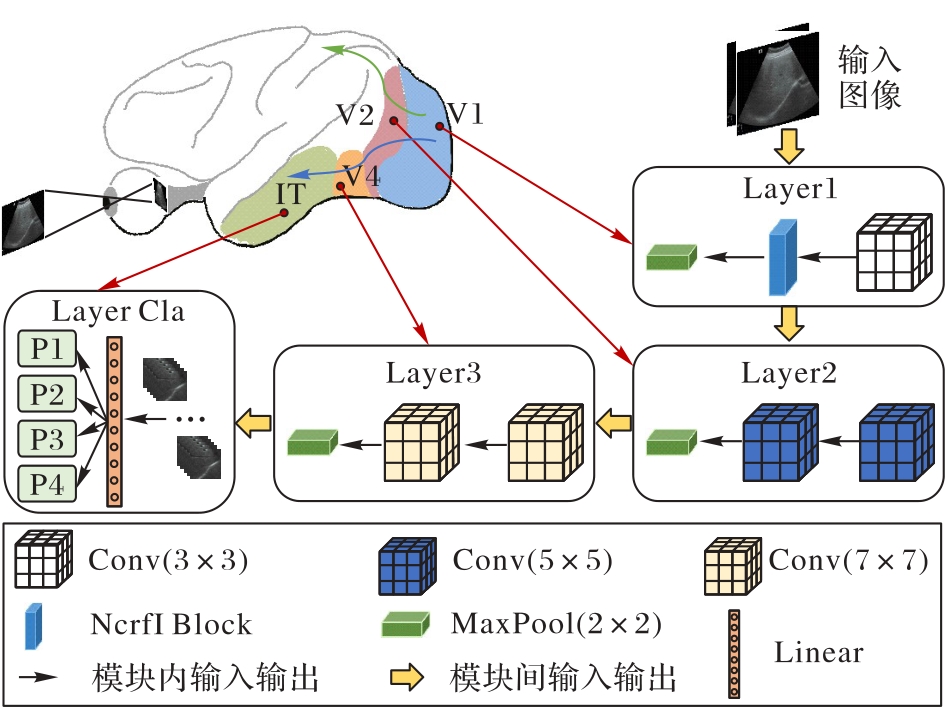
图1 VPNet的整体网络结构
Fig. 1 Overall network structure of VPNet
| 层 | 分支结构 | Parameter | |
|---|---|---|---|
| Layer1 | Conv(3×3) | (3,32,3×3,2) | |
| NcrfI Block | CRF Block | (32,64,3×3,2) | |
| nCRF Block | (64,64,5×5,1)-(64,64,3×3,1) | ||
| MaxPool | (64,64,1×1,2) | ||
| Layer2 | Conv(5×5) | (64,128,5×5,2) | |
| Conv(5×5) | (128,128,5×5,2) | ||
| MaxPool | (128,128,1×1,2) | ||
| Layer3 | Conv(7×7) | (128,256,7×7,2) | |
| Conv(7×7) | (256,256,7×7,2) | ||
| MaxPool | (256,256,1×1,2) | ||
| Layer Cla | Linear | featurein=2 304,featureout=4 | |
表1 VPNet的网络结构参数
Tab. 1 Network structure parameters of VPNet
| 层 | 分支结构 | Parameter | |
|---|---|---|---|
| Layer1 | Conv(3×3) | (3,32,3×3,2) | |
| NcrfI Block | CRF Block | (32,64,3×3,2) | |
| nCRF Block | (64,64,5×5,1)-(64,64,3×3,1) | ||
| MaxPool | (64,64,1×1,2) | ||
| Layer2 | Conv(5×5) | (64,128,5×5,2) | |
| Conv(5×5) | (128,128,5×5,2) | ||
| MaxPool | (128,128,1×1,2) | ||
| Layer3 | Conv(7×7) | (128,256,7×7,2) | |
| Conv(7×7) | (256,256,7×7,2) | ||
| MaxPool | (256,256,1×1,2) | ||
| Layer Cla | Linear | featurein=2 304,featureout=4 | |
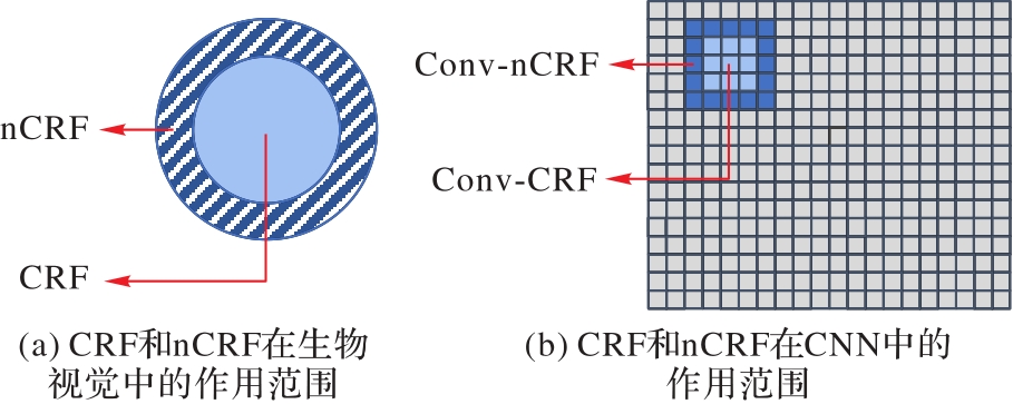
图2 CRF和nCRF的作用范围
Fig. 2 Action ranges of CRF and nCRF
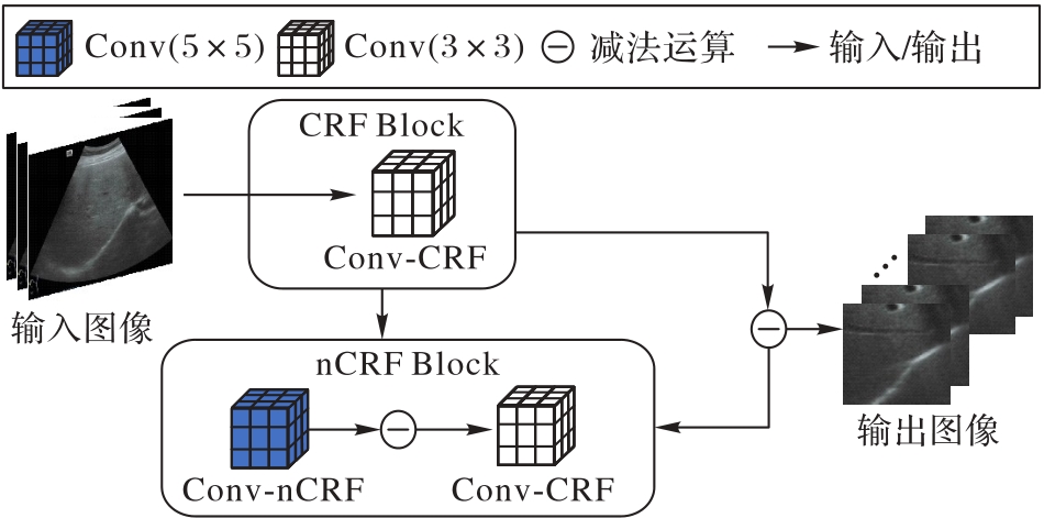
图3 NcrfI的网络结构
Fig. 3 Network structure of NcrfI
| 方法 | 准确率/% | WP | WR | WF1 |
|---|---|---|---|---|
| original | 74.42 | 0.79 | 0.74 | 0.75 |
| VPNet-VP | 81.40 | 0.82 | 0.81 | 0.81 |
| VPNet-NcrfI | 81.40 | 0.85 | 0.81 | 0.81 |
| VPNet | 88.37 | 0.89 | 0.88 | 0.88 |
表2 自制数据集FLUS-Datas上的消融实验结果
Tab. 2 Ablation experimental results on self-made dataset FLUS-Datas
| 方法 | 准确率/% | WP | WR | WF1 |
|---|---|---|---|---|
| original | 74.42 | 0.79 | 0.74 | 0.75 |
| VPNet-VP | 81.40 | 0.82 | 0.81 | 0.81 |
| VPNet-NcrfI | 81.40 | 0.85 | 0.81 | 0.81 |
| VPNet | 88.37 | 0.89 | 0.88 | 0.88 |
| 方法 | 准确率/% | WP | WR | WF1 |
|---|---|---|---|---|
| GLCM-SVM[ | 60.47 | 0.62 | 0.60 | 0.60 |
| InceptionResNetv2-SVM[ | 72.09 | 0.79 | 0.72 | 0.73 |
| ResNet101-SVM[ | 76.74 | 0.79 | 0.77 | 0.77 |
| 随机森林算法[ | 65.12 | 0.69 | 0.65 | 0.65 |
| EffficientNet[ | 76.74 | 0.79 | 0.77 | 0.77 |
| ViT[ | 69.77 | 0.72 | 0.67 | 0.65 |
| RepVGG[ | 79.07 | 0.80 | 0.79 | 0.79 |
| VPNet | 88.37 | 0.89 | 0.88 | 0.88 |
表3 自制数据集FLUS-Datas上的对比实验结果评估
Tab. 3 Evaluation of comparative experimental results on self-made dataset FLUS-Datas
| 方法 | 准确率/% | WP | WR | WF1 |
|---|---|---|---|---|
| GLCM-SVM[ | 60.47 | 0.62 | 0.60 | 0.60 |
| InceptionResNetv2-SVM[ | 72.09 | 0.79 | 0.72 | 0.73 |
| ResNet101-SVM[ | 76.74 | 0.79 | 0.77 | 0.77 |
| 随机森林算法[ | 65.12 | 0.69 | 0.65 | 0.65 |
| EffficientNet[ | 76.74 | 0.79 | 0.77 | 0.77 |
| ViT[ | 69.77 | 0.72 | 0.67 | 0.65 |
| RepVGG[ | 79.07 | 0.80 | 0.79 | 0.79 |
| VPNet | 88.37 | 0.89 | 0.88 | 0.88 |
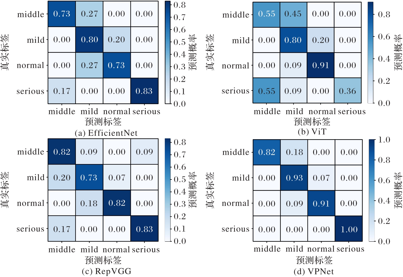
图4 FLUS-Datas数据集上4种方法的混淆矩阵对比
Fig. 4 Comparison of confusion matrices of four methods on FLUS-Datas dataset
| 方法 | Accuracy | Sensitivity | Specificity |
|---|---|---|---|
| GLCM-SVM[ | 85.4 | 84.2 | 88.2 |
| InceptionResNetv2-SVM[ | 96.3 | 100.0 | 88.2 |
| ResNet101-SVM[ | 99.3 | 98.6 | 100.0 |
| 随机森林算法[ | 100.0 | 100.0 | 100.0 |
| EfficientNet[ | 100.0 | 100.0 | 100.0 |
| ViT[ | 90.9 | 82.0 | 95.0 |
| RepVGG[ | 100.0 | 100.0 | 100.0 |
| VPNet | 100.0 | 100.0 | 100.0 |
表4 公开数据集上的对比实验结果评估 (%)
Tab. 4 Evaluation of comparative experimental results on public dataset
| 方法 | Accuracy | Sensitivity | Specificity |
|---|---|---|---|
| GLCM-SVM[ | 85.4 | 84.2 | 88.2 |
| InceptionResNetv2-SVM[ | 96.3 | 100.0 | 88.2 |
| ResNet101-SVM[ | 99.3 | 98.6 | 100.0 |
| 随机森林算法[ | 100.0 | 100.0 | 100.0 |
| EfficientNet[ | 100.0 | 100.0 | 100.0 |
| ViT[ | 90.9 | 82.0 | 95.0 |
| RepVGG[ | 100.0 | 100.0 | 100.0 |
| VPNet | 100.0 | 100.0 | 100.0 |
| 1 | LEE D H. Imaging evaluation of non-alcoholic fatty liver disease: focused on quantification[J]. Clinical and Molecular Hepatology, 2017, 23(4): 290-301. |
| 2 | YANG J, FAN J, AI D, et al. Local statistics and non-local mean filter for speckle noise reduction in medical ultrasound image[J]. Neurocomputing, 2016, 195: 88-95. |
| 3 | GUAN F, TON P, GE S, et al. Anisotropic diffusion filtering for ultrasound speckle reduction[J]. SCIENCE CHINA Technological Sciences, 2014, 57(3): 607-614. |
| 4 | BALOCCO S, GATTA C, PUJOL O, et al. SRBF: speckle reducing bilateral filtering[J]. Ultrasound in Medicine and Biology, 2010, 36(8): 1353-1363. |
| 5 | DAMODARAN N, RAMAMURTHY S, VELUSAMY S, et al. Speckle noise reduction in ultrasound biomedical B-scan images using discrete topological derivative[J]. Ultrasound in Medicine and Biology, 2012, 38(2): 276-286. |
| 6 | GAI S, ZHANG B, YANG C, et al. Speckle noise reduction in medical ultrasound image using monogenic wavelet and Laplace mixture distribution[J]. Digital Signal Processing, 2018, 72: 192-207. |
| 7 | RHYOU S Y, YOO J C. Cascaded deep learning neural network for automated liver steatosis diagnosis using ultrasound images[J]. Sensors, 2021, 21(16): No.5304. |
| 8 | ZHANG L, ZHU H, YANG T. Deep neural networks for fatty liver ultrasound images classification[C]// Proceedings of the 2019 Chinese Control and Decision Conference. Piscataway: IEEE, 2019: 4641-4646. |
| 9 | WU C H, HUNG C L, LEE T Y, et al. Fatty liver diagnosis using deep learning in ultrasound image[C]// Proceedings of the 2022 IEEE International Conference on Digital Health. Piscataway: IEEE, 2022: 185-192. |
| 10 | KARAGOZ M A, AKAY B, BASTURK A, et al. An unsupervised transfer learning model based on convolutional auto encoder for non-alcoholic steatohepatitis activity scoring and fibrosis staging of liver histopathological images[J]. Neural Computing and Applications, 2023, 35(14): 10605-10619. |
| 11 | BANZATO T, BONSEMBIANTE F, ARESU L, et al. Use of transfer learning to detect diffuse degenerative hepatic diseases from ultrasound images in dogs: a methodological study[J]. The Veterinary Journal, 2018, 233: 35-40. |
| 12 | BYRA M, STYCZYNSKI G, SZMIGIELSKI C, et al. Transfer learning with deep convolutional neural network for liver steatosis assessment in ultrasound images[J]. International Journal of Computer Assisted Radiology and Surgery, 2018, 13(12): 1895-1903. |
| 13 | SAHA S, SHEIKH N. Ultrasound image classification using ACGAN with small training dataset[C]// Proceedings of the 2020 International Symposium on Signal and Image Processing, AISC 1333. Singapore: Springer, 2021: 85-93. |
| 14 | 张顺,龚怡宏,王进军. 深度卷积神经网络的发展及其在计算机视觉领域的应用[J]. 计算机学报, 2019, 42(3):453-482. |
| ZHANG S, GONG Y H, WANG J J. Development of deep convolutional neural network and tts applications in computer vision[J]. Chinese Journal of Computers, 2019, 42(3): 453-482. | |
| 15 | CHE H, BROWN L G, FORAN D J, et al. Liver disease classification from ultrasound using multi-scale CNN[J]. International Journal of Computer Assisted Radiology and Surgery, 2021, 16(9): 1537-1548. |
| 16 | RIESENHUBER M, POGGIO T. Hierarchical models of object recognition in cortex[J]. Nature Neuroscience, 1999, 2(11):1019-1025. |
| 17 | 杨曦,闫杰,王文,等. 脑启发的视觉目标识别模型研究与展望[J]. 计算机工程与应用, 2022, 58(7):1-20. |
| YANG X, YAN J, WANG W, et al. Research and Prospect of brain-inspired models for visual object recognition[J]. Computer Engineering and Applications, 2022, 58(7): 1-20. | |
| 18 | ROLLS E T, MILWARD T. A model of invariant object recognition in the visual system: learning rules, activation functions, lateral inhibition, and information-based performance measures[J]. Neural Computation, 2000, 12(11): 2547-2572. |
| 19 | 姚行中,鲁统伟,胡汉平. 基于灵长类视觉皮层的目标识别模型综述[J]. 模式识别与人工智能, 2009, 22(4):581-588. |
| YAO X Z, LU T W, HU H P. Object recognition models based on primate visual cortex: a review[J]. Pattern Recognition and Artificial Intelligence, 2009, 22(4): 581-588. | |
| 20 | KRIZHEVSKY A, SUTSKEVER I, HINTON G E. ImageNet classification with deep convolutional neural networks[J]. Communications of the ACM, 2017, 60(6): 84-90. |
| 21 | LE Q V. Building high-level features using large scale unsupervised learning[C]// Proceedings of the 2013 IEEE International Conference on Acoustics, Speech and Signal Processing. Piscataway: IEEE, 2013: 8595-8598. |
| 22 | ZWEIG S, WOLF L. InterpoNet, a brain inspired neural network for optical flow dense interpolation[C]// Proceedings of the 2017 IEEE Conference on Computer Vision and Pattern Recognition. Piscataway: IEEE, 2017: 6363-6372. |
| 23 | YU C P, LIU H, SAMARAS D, et al. Modelling attention control using a convolutional neural network designed after the ventral visual pathway[J]. Visual Cognition, 2019, 27(5/6/7/8): 416-434. |
| 24 | KARIMI-ROUZBAHANI H, BAGHERI N, EBRAHIMPOUR R. Invariant object recognition is a personalized selection of invariant features in humans, not simply explained by hierarchical feed-forward vision models[J]. Scientific Reports, 2017, 7: No.14402. |
| 25 | 田媚,罗四维,齐英剑,等. 基于视觉系统“What”和“Where”通路的图像显著区域检测[J]. 模式识别与人工智能, 2006, 19(2):155-160. |
| TIAN M, LUO S W, QI Y J, et al. Detecting salient regions based on “What” and “Where” pathways of visual systems[J]. Pattern Recognition and Artificial Intelligence, 2006, 19(2): 155-160. | |
| 26 | NOTHDURFT H C, GALLANT J L, VAN ESSEN D C. Response modulation by texture surround in primate area V1: correlates of “popout” under anesthesia[J]. Visual Neuroscience, 1999, 16(1): 15-34. |
| 27 | DENG S, LIU N, HUO H, et al. Contour detection based on multi-scale spatial inhibition and contextual modulation[C]// Proceedings of the 7th International Conference on Advanced Computational Intelligence. Piscataway: IEEE, 2015: 372-377. |
| 28 | GOODALE M A, MILNER A D. Separate visual pathways for perception and action[J]. Trends in Neurosciences, 1992, 15(1): 20-25. |
| 29 | KAR K, KUBILIUS J, SCHMIDT K, et al. Evidence that recurrent circuits are critical to the ventral stream’s execution of core object recognition behavior[J]. Nature Neuroscience, 2019, 22(6): 974-983. |
| 30 | FIEHLER K, BURKE M, BIEN S, et al. The human dorsal action control system develops in the absence of vision[J]. Cerebral Cortex, 2009, 19(1): 1-12. |
| 31 | 孙莹,孙洵伟,王一帆,等. 跨通道迁移及其认知神经机制[J]. 生物化学与生物物理进展, 2024, 51(1):94-110. |
| SUN Y, SUN X W, WANG Y F, et al. Crossmodal transfer and its cognitive neural mechanisms[J]. Progress in Biochemistry and Biophysics, 2024, 51(1): 94-110. | |
| 32 | 林点,潘理,易平. 面向图像识别的卷积神经网络鲁棒性研究进展[J]. 网络与信息安全学报, 2022, 8(3):111-122. |
| LIN D, PAN L, YI P. Research on the robustness of convolutional neural networks in image recognition[J]. Chinese Journal of Network and Information Security, 2022, 8(3): 111-122. | |
| 33 | 李冰. 灵长类视觉目标识别的神经机制研究进展[J]. 中国药理学与毒理学杂志, 2017, 31(11):1057-1062. |
| LI B. Mechanisms underlying visual object recognition in primates: research progress[J]. Chinese Journal of Pharmacology and Toxicology, 2017, 31(11): 1057-1062. | |
| 34 | CADIEU C, KOUH M, PASUPATHY A, et al. A model of V4 shape selectivity and invariance[J]. Journal of Neurophysiology, 2007, 98(3): 1733-1750. |
| 35 | 王伟锋,金杰,陈景明. 基于感受野的快速小目标检测算法[J]. 激光与光电子学进展, 2020, 57(2): No.021501. |
| WANG W F, JIN J, CHEN J M. Rapid detection algorithm for small objects based on receptive field block[J]. Laser and Optoelectronics Progress, 2020, 57(2): No.021501. | |
| 36 | 郑雅菁,余肇飞,黄铁军. 生物视觉系统的神经网络编码模型综述[J]. 中国图象图形学报, 2023, 28(2):335-357. |
| ZHENG Y J, YU Z F, HUANG T J. A literature review for neural networks-based encoding models of biological visual system[J]. Journal of Image and Graphics, 2023, 28(2): 335-357. | |
| 37 | HASANI H, BAGHSHAH M S, AGHAJAN H. Surround modulation: a bio-inspired connectivity structure for convolutional neural networks[C]// Proceedings of the 33rd International Conference on Neural Information Processing Systems. Red Hook: Curran Associates Inc., 2019: 15903-15914. |
| 38 | 窦燕,康锦华,王丽盼. 结合人眼微动的新型非经典感受野模型[J]. 光学学报, 2019, 39(3): No.0310002. |
| DOU Y, KANG J H, WANG L P. Novel non-classical receptive field model combined with human eye fretting[J]. Acta Optica Sinica, 2019, 39(3): No.0310002. | |
| 39 | 覃溪,李兴. 非经典感受野的轮廓检测模型研究[J]. 中阿科技论坛(中英文), 2020(9):120-123. |
| QING X, LI X. Contour detection model based on non-classical receptive field[J]. China-Arab Science and Technology Forum, 2020(9): 120-123. | |
| 40 | LI C Y, LI W. Extensive integration field beyond the classical receptive field of cat’s striate cortical neurons — classification and tuning properties[J]. Vision Research, 1994, 34(18): 2337-2355. |
| 41 | GRIGORESCU C, PETKOV N, WESTENBERG M A. Contour detection based on nonclassical receptive field inhibition[J]. IEEE Transactions on Image Processing, 2003, 12(7): 729-739. |
| 42 | TANG Q, SANG N, LIU H. Contrast-dependent surround suppression models for contour detection[J]. Pattern Recognition, 2016, 60: 51-61. |
| 43 | TANG Q, SANG N, LIU H. Learning nonclassical receptive field modulation for contour detection[J]. IEEE Transactions on Image Processing, 2020, 29: 1192-1203. |
| 44 | TAN M, LE Q V. EfficientNet: rethinking model scaling for convolutional neural networks[C]// Proceedings of the 36th International Conference on Machine Learning. New York: JMLR.org, 2019: 6105-6114. |
| 45 | DOSOVITSKIY A, BEYER L, KOLESNIKOV A, et al. An image is worth 16×16 words: Transformers for image recognition at scale[EB/OL]. [2023-10-22].. |
| 46 | DING X, ZHANG X, MA N, et al. RepVGG: making VGG-style ConvNets great again[C]// Proceedings of the 2021 IEEE/CVF Conference on Computer Vision and Pattern Recognition. Piscataway: IEEE, 2021: 13728-13737. |
| 47 | HO T K. Random decision forests[C]// Proceedings of the 3rd International Conference on Document Analysis and Recognition — Volume 1. Piscataway: IEEE, 1995: 278-282. |
| 48 | ZAMANIAN H, MOSTAAR A, AZADEH P, et al. Implementation of combinational deep learning algorithm for non-alcoholic fatty liver classification in ultrasound images[J]. Journal of Biomedical Physics and Engineering, 2021, 11(1): 73-84. |
| [1] | 邓淼磊, 阚雨培, 孙川川, 徐海航, 樊少珺, 周鑫. 基于深度学习的网络入侵检测系统综述[J]. 《计算机应用》唯一官方网站, 2025, 45(2): 453-466. |
| [2] | 余松森, 林智凡, 薛国鹏, 徐建宇. 基于改进YOLOv8的轻量级大幅面瓷砖缺陷检测算法[J]. 《计算机应用》唯一官方网站, 2025, 45(2): 647-654. |
| [3] | 张天骐, 谭霜, 沈夕文, 唐娟. 融合注意力机制和多尺度特征的图像水印方法[J]. 《计算机应用》唯一官方网站, 2025, 45(2): 616-623. |
| [4] | 洪梓榕, 包广清. 基于集成学习的雷达自动目标识别综述[J]. 《计算机应用》唯一官方网站, 2025, 45(2): 371-382. |
| [5] | 张众维, 王俊, 刘树东, 王志恒. 多尺度特征融合与加权框融合的遥感图像目标检测[J]. 《计算机应用》唯一官方网站, 2025, 45(2): 633-639. |
| [6] | 张思齐, 张金俊, 王天一, 秦小林. 基于信号时态逻辑的深度时序事件检测算法[J]. 《计算机应用》唯一官方网站, 2025, 45(1): 90-97. |
| [7] | 郑宗生, 杜嘉, 成雨荷, 赵泽骋, 张月维, 王绪龙. 用于红外-可见光图像分类的跨模态双流交替交互网络[J]. 《计算机应用》唯一官方网站, 2025, 45(1): 275-283. |
| [8] | 徐欣然, 张绍兵, 成苗, 张洋, 曾尚. 基于多路层次化混合专家模型的轴承故障诊断方法[J]. 《计算机应用》唯一官方网站, 2025, 45(1): 59-68. |
| [9] | 梁杰涛, 罗兵, 付兰慧, 常青玲, 李楠楠, 易宁波, 冯其, 何鑫, 邓辅秦. 基于坐标几何采样的点云配准方法[J]. 《计算机应用》唯一官方网站, 2025, 45(1): 214-222. |
| [10] | 晏燕, 钱星颖, 闫鹏斌, 杨杰. 位置大数据的联邦学习统计预测与差分隐私保护方法[J]. 《计算机应用》唯一官方网站, 2025, 45(1): 127-135. |
| [11] | 秦璟, 秦志光, 李发礼, 彭悦恒. 基于概率稀疏自注意力神经网络的重性抑郁疾患诊断[J]. 《计算机应用》唯一官方网站, 2024, 44(9): 2970-2974. |
| [12] | 王熙源, 张战成, 徐少康, 张宝成, 罗晓清, 胡伏原. 面向手术导航3D/2D配准的无监督跨域迁移网络[J]. 《计算机应用》唯一官方网站, 2024, 44(9): 2911-2918. |
| [13] | 黄云川, 江永全, 黄骏涛, 杨燕. 基于元图同构网络的分子毒性预测[J]. 《计算机应用》唯一官方网站, 2024, 44(9): 2964-2969. |
| [14] | 潘烨新, 杨哲. 基于多级特征双向融合的小目标检测优化模型[J]. 《计算机应用》唯一官方网站, 2024, 44(9): 2871-2877. |
| [15] | 李顺勇, 李师毅, 胥瑞, 赵兴旺. 基于自注意力融合的不完整多视图聚类算法[J]. 《计算机应用》唯一官方网站, 2024, 44(9): 2696-2703. |
| 阅读次数 | ||||||
|
全文 |
|
|||||
|
摘要 |
|
|||||